- Visibility 88 Views
- Downloads 8 Downloads
- DOI 10.18231/j.ijpo.2023.022
-
CrossMark

Vulvar swelling- A rare diagnosis
- Author Details:
-
 Ahmad Raza *
-
Sumaiya Irfan
-
Noorin Zaidi
-
Nirupma Lal
Introduction
Breast pathologies require recognition because of its high incidence and prevalence, influence on women's life and its premalignant potential. One of the most common benign pathologies of the Breast is Fibroadenoma which is a fibroepithelial lesion. It generally occurs in the breast but since the milk line is present in many parts of the body, mammary-like glands can also be found in between the sulcus of labium minora and labium majora. These glands are made up of ducts, with accompanying acini, and are lined with breast-like glandular epithelium. These glands' draining channels exit through the skin. Many of the physiologic and pathologic changes that affect the normal breast parenchyma, also affect the ectopic breast tissue. The clinical features of vulvar fibroadenoma include cyclic pain during menstruation, swelling, lactation during pregnancy, etc. It may complicate to form cysts, pseudoangiomatous stromal hyperplasia (PASH), Papillary hidradenoma, carcinoma, etc. Hereby, we are presenting a case of vulvar fibroadenoma in a 45 years old woman.
Case Report
A 45-year-old female presented to the gynecology opd with the chief complaints of painless, well-defined mass in the left labia majora for one year which was gradually progressive. On physical examination, the size of the swelling was 4x2cm, smooth, firm, mobile and non tender. Her general and systemic examination was normal. All the hematological as well as biochemical investigations were within normal limits. On radiological examination, an ultrasonography showed evidence of a well-defined rounded heterogeneously hypoechoic space occupying lesion measuring ~3.5x2.5x1cm noted in the left labia majora suggestive of fibroadenoma. Thus, the lesion was excised under Local Anesthesia. Then the specimen was sent to histopathology laboratory for further processing.
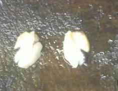
Gross
A single gray-white to gray-brown, smooth soft tissue piece measuring 3x2x1cm was received at our histopathology department. Cut surface revealed a gray-white solid areas. Representative sections were taken, then tissue was processed and routine H&E staining was done.
Microscopy
Light microscopic examination showed breast tissue with proliferation of cystically dilated glands and stromal elements. These glands showed predominantly intracanalicular patterns in which connective tissues invaginated into glandular spaces so that it appeared to be within them which were lined by low columnar to cuboidal epithelium surrounded by the outer layer of myoepithelial cells. There was also the presence of pericanalicular pattern in which the ducts were round to oval in shape and were lined by inner layer of cuboidal epithelium and outer layer of myoepithelial cells. The intervening stroma was myxomatous.


Histopathologic impression of vulvar fibroadenoma was made.
Discussion
The epidemiology and ethnic predisposition of vulvar fibroadenoma is understood poorly. Few studies suggested its occurrence in 1-6% of the population.[1], [2]
The pathogenesis of vulvar fibroadenoma is debatable and unsure. One theory states that primitive embryological milk line gives rise to ectopic mammary tissue which will finally form vulvar fibroadenoma. Incomplete involution of these mammary tissues lead to the formation of vulvar fibroadenoma.[3], [1], [2], [4] Another study suggested that there are specialized glands similar to mammary glands present in the genital areas. These glands are known as mammary-like anogenital glands which give rise to vulvar fibroadenoma.[1], [2], [4], [5]
The vulval fibroadenoma may present as painful or asymptomatic swelling which may show variation during pregnancy and lactation, cyclic pain, etc.[3], [1], [2], [4] Generally it occurs in females between the ages of 18-84 years.[6] It may also complicate to form cysts, pseudoangiomatous stromal hyperplasia (PASH), Papillary hidradenoma, carcinoma, etc. In our case, a 45 years old female presented with painless, well circumscribed mass in the vulva and no complications were observed.
Some of the benign differential diagnosis of this pathology are epidermal cyst, follicular cyst, Bartholin’s gland duct cyst, lipoma, hidradenoma papilliferum, lactating adenoma, intraductal papilloma, apocrine adenoma, phyllodes, syringoma, pseudoangiomatous stromal hyperplasia, fibrocystic disease and sclerosing adenosis.[7] Few malignant differentials are extramammary Paget’s disease, ductal/lobular/mucinous adenocarcinoma.[7]
The vulvar fibroadenomas can be diagnosed using fine needle aspiration cytology, Ultrasonography and histopathological examination. Histopathological examination is similar to the breast fibroadenoma, having proliferation of cystically dilated glands and stromal elements with intracanalicular and pericanalicular patterns. The glands are lined by the inner layer of cuboidal epithelium and outer layer of myoepithelial cells.
Immunohistochemistry can also be used in the diagnosis. Some of the markers are ER, PR, CK, S100, SMA, EMA and GCDF-15.
The mainstay treatment of this condition is surgery. Excision usually has a good prognosis and rarely recurs. After 6months of follow up, our patient was doing well and there was no evidence of recurrence.
Conclusion
Although vulvar fibroadenoma is a rare finding, we should keep it as a differential diagnosis in cases of vulvar mass or pedunculated soft tissue mass. It is necessary to promote awareness regarding vulvar fibroadenoma.
Ethics Approval
Ethical approval obtained from institutional ethics committee of Era’s Lucknow Medical College & Hospital.
Consent for Publication
Consent was given by the patient.
Source of Funding
No source of funding.
Authors Contribution
All authors contributed equally in the preparation of manuscript.
Conflict of Interest
None.
Acknowledgement
None.
References
- DC DeLeon, DP Montiel, H Vázquez, C Hernández, L Cetina, MH Lucio. Vulvar fibroadenoma: a common neoplasm in an uncommon site. World J Surg Oncol 2009. [Google Scholar]
- CC Anunibi, FJN Obiajulu, AAF Banjo, AOE Okonkwo. Vulval fibroadenoma associated with lactating adenoma in a 26 year old Nigerian female. Case Rep Pathol 2013. [Google Scholar]
- AZ mirzaie, MTH Ashtiani, B Kazeminejad. Fibroadenoma of the vulva: a case report. Iran J Pathol 2008. [Google Scholar]
- WC Lin, WL Lin, YH Chuang, PY Shih, JC Lee, HS Hong. An asymptomatic nodule in the vulva. Clin Exp Dermatol 2008. [Google Scholar]
- PD Maj, D Swarup, GC Mishra. Fibroadenoma of aberrant breast tissue in the vulva. Med J Armed Forces India 2000. [Google Scholar]
- AL Lopez, S Manandhar, L Dubow. Pregnancy-unrelated fibroadenoma in ectopic breast tissue in the axilla and vulva: A case report. Case Rep Womens Health 2020. [Google Scholar]
- R Kalyani, MV Srinivas, P Veda. Vulval fibroadenoma-a report of two cases with review of literature. Int J Biomed Sci 2014. [Google Scholar]