Introduction
Kimura’s disease is an inflammatory disorder of unknown etiology.1 It is seen in an endemic form in Asia but it is also present in other parts of the world.2 Kimura’s disease typically manifests with subcutaneous nodules in the regions of head and neck, often as unilateral swelling and commonly accompanied by regional lymphnodes enlargement. It is sometimes involves salivary glands. Kimura’s disease also associated with increase level of eosinophils in the blood and tissue along with increase IgE levels.3, 4, 5 Kimura's disease is uncommon in India, with only around 200 cases reported globally. Given its rarity, accurate histopathological diagnosis is crucial for proper management.6 It is Typically observed in young adults, Kimura’s disease predominantly affects individuals aged between 20 and 40 years, with a higher incidence in men, with a ratio of 3:1 compared to women.7, 2 The frequently involved sites in Kimura’s disease are periauricular region, eyelids, orbital regions and also in groin area.4 Renal disease often coexists with Kimura’s disease, with reported incidence rates ranging from 10% to 60%.2 Diagnosis of Kimura’s disease can be challenging, and it typically requires a biopsy or excision of the affected swelling for histopathological analysis to confirm the diagnosis.
Case History
A male patient with age of 26 year came to our hospital with history of post auricular swelling for two years which was progressively increase in size and sudden in onset. There was no history of any other symptoms like fever, weight loss etc. Overlying skin of swelling was normal. FNAC was performed, findings were suggestive of Lymph node hyperplasia with eosinophilia – Suggestive of Kimura’s disease. CECT findings shows Homogenously enhancing soft tissue density lesion in skin and subcutaneous plane of post auricular region which suggest possibility of benign soft tissue lesion likely. Excision biopsy was advised. Left post auricular swelling excision was done and tissue sent for histopathological examination.
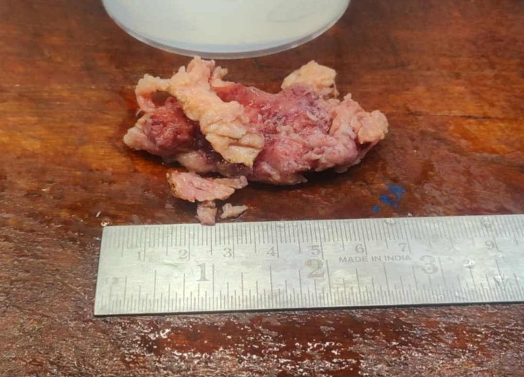
Gross examination
Specimen consists of multiple reddish whitish soft tissue portions, total measuring 6.8 × 4.2 cm. The outer surface of the received specimen appeared irregular, while the cut surface exhibited a whitish, homogeneous appearance.
Microscopic examination
Histopathological Sections reveal Lymphnodes with preserved but distorted overall architecture with hyperplastic follicles with germinal centres associated with expansion of interfollicular areas by abundant eosinophils with presence of few areas of eosinophilic micro abscess are seen. The examination revealed vascular proliferation and a polymorphous population consisting of many lymphocytes, plasma cells, histiocytes, and numerous eosinophils. Focal areas of sclerosis were also present. Histological features were suggestive of Kimura’s disease.
Figure 2
Lymphoid follicles which are hyperplastic and also present of reactive germinal centers (10x)


Discussion
Kimura's disease, also referred to as eosinophilic granuloma. Typically, Kimura’s disease presents with a triad of 1) peripheral eosinophilia 2) increased level of IgE and 3) one or more subcutaneous indolent nodules.8 The Exact pathogenesis and etiology of Kimura’s disease are remained unclear. It is speculated to involve a self-limited allergic or autoimmune response triggered by an unknown persistent antigenic stimulus.3
Kimura’s disease present as a subcutaneous swelling in the head and neck region. Sometimes as a mass lesion in the major salivary glands and often associated with regional lymph node enlargement. Sometimes patient presented with only enlargement of lymphnodes.9 Microscopically, the involved nodes show marked hyperplasia of germinal centers, a few of which may be of the progressively transformed type. These germinal centers are often well vascularized and contain polykaryocytes, interstitial fibrosis, and deposition of a proteinaceous material. There is also extensive infiltration by mature eosinophils, with occasional formation of eosinophilic abscesses. Hyalinized vessels are often seen in the paracortical region, and there is a variable degree of sinus and paracortical sclerosis. An increase in the number of plasma cells and mast cells has been noted in the paracortex, together with proliferation of postcapillary venules. Some cases are associated with increases in IgG4-positive plasma cells, a feature of unclear significance.3, 5, 10
Angio lymphoid hyperplasia with eosinophilia (ALHE), Reactive follicular hyperplasia, parasitic lymphadenitis, Castleman’s disease, tuberculosis, Hodgkin’s disease (mixed cellularity type), Kaposi sarcoma and Langerhans cell histiocytosis all are differential diagnosis of Kimura’s disease.5 Current evidence strongly suggests that Kimura’s disease and the disease known to dermatologists as Angio lymphoid hyperplasia (ALHE) with eosinophilia are different entities. Both entities show microscopically vascular proliferations and infiltrations of eosinophils. Vascular proliferations are more significant in Angio lymphoid hyperplasia with eosinophilia. ALHE is more common in western population, middle age females. Serum IgE levels are normal in Angio lymphoid hyperplasia with eosinophilia.
Kimura’s disease Histologically exhibits three components: 1) cellular components characterised by inflammatory infiltrate with increased eosinophils and hyperplasia of lymph node follicles. 2) fibrocollagenous component and 3) vascular proliferation of the postcapillary venule.3, 11 Other possible conditions to consider when diagnosing Kimura’s disease affecting the parotid gland include Mikulicz's disease, infective parotitis, Sjogren's syndrome, and different types of salivary gland tumors.12
Kimura’s disease usually follows a benign and self-limiting clinical course. Patients often experience a prolonged period with gradual swelling enlargement, occasionally witnessing spontaneous resolution.3
Imaging studies play a crucial role in diagnosis and can assist in staging the extent and progression of Kimura’s disease, including evaluating lymph node involvement. Treatment options for Kimura’s disease include surgical excision, steroid therapy and radiation.3
Conclusion
Kimura’s disease should be considered as a potential diagnosis in patients presenting with head and neck masses and lymphadenopathy, and investigations should be carried out accordingly. Henceforth, histopathological examination plays very important roll to diagnose Kimura’s disease. Understanding Kimura’s disease will enable clinicians to make more accurate diagnoses and provide appropriate treatment to the patients.