Introduction
Florid vascular proliferation is a rare entity in the gastrointestinal tract, seen most commonly secondary to obstruction. Although this is a benign and reactive process, it can mimic vascular tumours clinically as well as histologically.1, 2 Pathologist needs to be aware of the pathogenesis and histology of this lesion, so a misdiagnosis of vascular neoplasm can be avoided. Here we report a case with florid vascular proliferation in the colon, secondary to intussusception caused due to a large tubulo-villous adenoma transforming to adenocarcinoma.
Case History
A 47-year-old man presented with abdominal discomfort and vomiting. Computed Tomography and Ultrasonography suggested ileo-colic intussusception. Colonoscopy revealed a polypoidal mass in the ascending colon and the biopsy from the mass showed villous adenoma with high grade dysplasia. Invasion was not seen.
Three days later, surgical intervention was performed and excision of intussuscepted bowel from distal ileum to colon distal to the mass was done. Histopathological examination of colon revealed a large polypoidal growth measuring 6 x 5.5cm with nodular and fleshy cut surface. The mucosa adjacent to the mass was thickened and showed a granular, friable appearance.
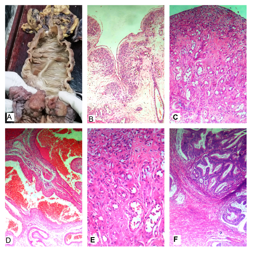
Several sections studied through the mass revealed a well differentiated adenocarcinoma arising in a tubulo-villous adenoma with high grade dysplasia. The tumour infiltrated the submucosal layer and there was no evidence of lymphovascular / perineural invasion. There was extensive vascular proliferation most evident in the section studied through the granular appearing bowel wall, adjacent to the tumour. Histologically, the mucosa was thrown into lobular pattern with numerous small and large capillaries, crowded throughout the mucosa and submucosa, extending into the serosa with focal areas of haemorrhages. Many large ectatic and congested blood vessels were also seen.
Figure 1
a): Gross photo of Hemicolectomy specimen showing polypoidal growth and shiny greyish lesion noted; b): Lobular proliferation of capillary sized vasculature, H&E stain, 40X; c): Lobular proliferation of capillary sized vasculature, H&E stain, 40X; d): Large irregular dilated & congested vascular channels, H&E stain, 200X; e): Lobular proliferation of capillary sized vasculature, H&E stain, 200X; f): Tubulo-villous adenoma with foci of invasion into submucosa, H&E stain, 200X.

Discussion
Intussusception is relatively rare in adult age group accounting to 5% of overall cases.3, 4 The most common causes of intussusception include tumours, lipoma or polyps in adults and Meckel’s diverticulum in children.4 Florid vascular proliferation in the bowel in association with intussusception is an enigmatic entity.3, 4
Intussusception leads to several reactive changes in the bowel like mucosal ulceration/ hyperplasia, disorganisation of muscularis propria, telangiectasia, submucosal fibrosis, neural hyperplasia and serous adhesion. In addition to these, the mechanical stress resulting in mucosal prolapse could lead to bowel wall hypoperfusion.5, 6 As a response to hypoperfuion, angiogenesis is initiated by the nitric oxide and VEGF-A,7 resulting in proliferation of capillaries.
Gross examination findings in our case showed a polypoid mass with a fleshy, conglomerated and nodular appearance. Adjacent mucosa was thickened with a friable granular appearance. Several sections that were sampled showed FVP and foci of invasive adenocarcinoma were very few.
Microscopically, most FVPs show a lobular arrangement of capillary sized vascular channels lined by plump endothelial cells. There were several large thick walled vessels at the base of the lobules. The lesion mimics a lobular capillary haemangioma. Apart from this typical finding, vascular channels of varying calibres were seen involving the entire thickness of the bowel wall, extending from submucosa through the muscularis propria, into the subserosal adipose tissue. There was no evidence of nuclear atypia or conspicuous mitosis. The overlying mucosa showed complete loss of the mucosal crypt epithelial cells and was replaced by a flattened lining, beneath which the capillary proliferation was noted.
There are several entities that have to be considered in the differential diagnosis, in view of the extensive vascular proliferation. The list includes angiodysplasia, Kaposi sarcoma, and pyogenic granuloma. Lack of cellular atypia, absence of mitosis or necrosis and proliferation of spindle cells helps differentiate FVP from vascular neoplasms.5, 6
Angiodysplasia commonly involves small/ large intestine and histologically characterised by presence of thin walled vascular channels which tend to be dilated, vary in size, and extend primarily from submucosa into the mucosa.1 Kaposi sarcoma predominantly involves submucosa showing dense proliferation of spindle cells with nuclear atypia and intervening blood filled channels.1 Pyogenic granuloma histologically shows lobular pattern, with protuberant growth of capillary sized blood vessels lined by endothelial cells involving entire thickness of bowel wall.4, 5, 6
To conclude, florid vascular proliferation is common in the bowel, especially secondary to intussusceptions. The mechanism leading to vascular proliferation is hypoxia induced neo-angiogenesis. Pathologists have to be aware of this reactive proliferation of vessels in any case of intussusception, so that a mis-diagnosis of vascular tumour can be avoided.
