Introduction
Occurrence of benign mucinous metaplasia in the genital area is rare. A unique case of benign mucinous metaplasia of the prepuce in a 60-year-old man is reported here, so as to avoid over diagnosis of reactive nature of the lesion as neoplastic as its differentials being malignant entities like superficial spreading malignant melanoma, extramammary Paget's disease, mucinous syringometaplasia and cutaneous squamous cell carcinoma in situ with mucinous metaplasia.1
Clinical and pathological findings
60-year-old gentle man presented with complaints of phimosis, underwent circumcision and sample was received in the department of histopathology. Which was two skin covered soft tissue with no significant gross pathology.
Microscopy
Histopathology revealed mildly hyperkeratotic, irregularly acanthothic stratified squamous epithelium with dissipated mucin containing cells in the upper layers of the epithelium. Lamina propria shows moderate chronic inflammatory cell infiltrate and there was no evidence of dysplasia/malignancy in the areas analyzed.
Figure 1
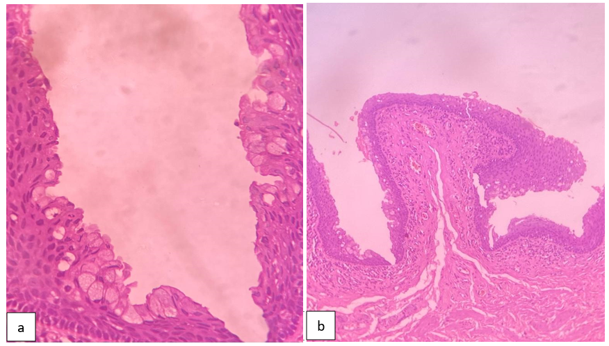
A,B: Sections show preputial skin with scattered mucin filled epithelium in superficial layers (H&E)
Alcian PAS and Periodic acid schiff was performed to affirm the presence of mucin and its nature, Cells with mucin in the epithelium are featured by Alcian PAS and Periodic acid schiff.

Discussion
Metaplasia is change in cell differentiation. One grown-up cell type is supplanted by another. Preputial skin don't have mucinous cells. Squamous cells goes through metaplastic change in upper layers of polygonal skin having columnar cells with multivacuolated mucin containing cytoplasm.2
Mucinous metaplasia is seen for different conditions such as mucinous syringometaplasia,3 extramammary Paget's disease,4 cutaneous in situ and intrusive squamous cell carcinoma with mucinous metaplasia, epidermotropic metastasis and mucinous papulosis.5
The criteria utilized for recognizing harmless mucinous metaplasia from various differentials are 1) The presence of benign and basally positioned nuclei 2) The metaplastic cells are found in the upper layers of the epithelium and the metaplastic cells are not infiltrative in nature rather it replaces the ordinary epithelium 3) The metaplastic cells are restricted to the epidermis.
Mucinous metaplasia is found in upper layers of epithelium. The metaplastic cells are frequently cuboidal or polygonal. The Spectrum changes in mucinous metaplasia is distinct, hence it should be differentiated from its differentials.6
Aw Fang et al., retrieved 100 prepuce example from the histopathology archives at Southampton General Hospital. Altogether, four instances of mucinous metaplasia were identified, one involving the glans penis and three involving the prepuce. All cases are seen between the age gathering of 51-80 years. One case showed non-specific balanitis and three cases were related with Zoon's balanitis.7
Mucinous metaplasia is reported in different genital regions like prepuce, vagina, vulva and glans penis. However, the pathogenesis of mucinous metaplasia of the genital region stays obscure. Mucinous metaplasia is seen associated with lichen sclerosus which is probably a chance or due to chronic inflammation.2