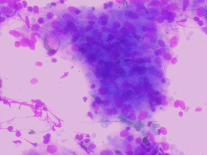
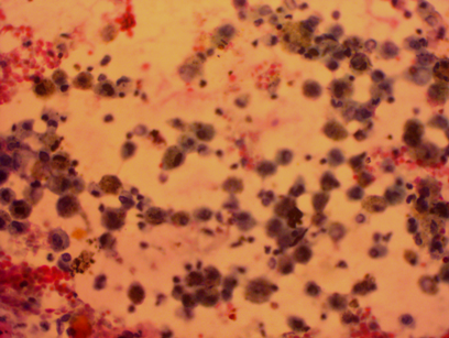
Introduction
The field of lung pathology is making rapid advances with better documentation of morphological changes in bronchial epithelium and other cells so that the earliest change from normal to diseased condition can be detected thus providing an early and accurate diagnosis to these non- neoplastic and neoplastic conditions of lung which will change the management.
Techniques like bronchial brushings (BB), bronchoalveolar lavage (BAL) and trans-bronchial needle aspiration (TBNA) became popular tools for obtaining diagnostic cytological material from various sites of the tracheo-bronchial passage. Today these cytological procedures constitute the most useful and least expensive investigative tools available for the detection of pulmonary diseases.1, 2, 3
Respiratory tract cytology is well established throughout the world as a diagnostic procedure in the evaluation of patient with suspected lung lesion.4, 5
The diagnostic and therapeutic efficacy of procedure depends on expertise, accessibility, condition of patient, site of lesion and other factors.
The most important utility of these procedures is in early diagnosis of neoplastic lesions as lung cancer is leading cause of cancer death worldwide.6 The most significant factor for survival in lung cancer is the stage of disease at diagnosis.7
Even in diagnosing non-neoplastic lesions like tuberculosis having wide spectrum of presentation bronchoscopic cytological modalities are extremely useful in confirming the diagnosis. Based on this background, this study was conducted as an attempt to compare efficacy of these bronchoscopic cytological modalities BAL, BB and TBNA with trans-bronchial lung biopsy (TBLB) as gold standard. Pulmonary lesions may be correctly diagnosed if multiple techniques are used to acquire diagnostic material.8
Aim & Objectives
The aim of this study is an attempt to compare the efficacy of cytological investigations (BAL, BB, and TBNA) in diagnosing lesions of lung, and comparing its result with gold standard, TBLB.
Materials and Methods
This was a cross-sectional, descriptive, observational study that was carried out in a tertiary care Hospital in Delhi over a period of two years. The sample size was calculated as per prevailing system. Informed consent of the patients were obtained and study was conducted on a heterogeneous dichotomous population. 200 patients were subjected cytology and biopsy of lungs and findings analyzed. Institutional ethical committee clearance was availed.
Cytological studies of BAL, BB, TBNA were done during routine diagnostic bronchoscopies of clinically diagnosed / suspected cases of lung lesions at our centre and their results were compared with TBLB as gold standard. Only the cases where BAL, BB, TBNA and TBLB were received simultaneously were included.
Preparation and collection of smears were done as per standard operating procedures. In each cases BAL, TBNA, BB and TBLB specimens were taken. The samples were also sent for Gram stain, bacterial culture and sensitivity, (Acid Fast Bacilli) AFB stain, Grocott / Periodic -Acid Schiff stain (PAS) stain and culture to microbiology department, if indicated.
Exclusion criteria
The paediatric age groups (< 18 years were excluded from the study.
Patients who had lung pathologies but no previous biopsy samples collected were also excluded.
Autolysed specimens with disturbed cellular morphological details or technical flaws were not included.
Cytological specimen analyzed for
Adequacy of sample.
Documentation of findings in epithelial cells.
Presence of any increase in acute inflammatory cells or persistence of chronic infection.
Presence of any malignancy.
All the slides are thoroughly screened with light microscope and criteria used to call sample as inadequate is as following:4, 5
Had fewer than 10 alveolar macrophages per high power field. As fewer alveolar macrophages does not represent true lung cytology.
Contain excessive numbers of epithelial cells, either showing morphologic degenerative changes or exceeding the number of alveolar macrophages present (>5%). As these will obscure the other cellular details.
Contain a mucopurulent exudate of polymorphonuclear cells obscuring lesion cellular features.
Contain excessive red blood cells due to trauma during the procedure.
Contain degenerative changes or laboratory artifacts obscuring cell identity, thus distorting the cytological details of lesion.
The biopsy specimen was processed in an automatic tissue processor for paraffin block preparation. From each block, 3 micron thick sections were cut by using Leica rotary microtome. All the slides were then stained with routine H & E staining methods. All the slides are thoroughly screened and the diagnosis was confirmed.
Histology slides were studied for:
Representation of adequate biopsy.
Morphological findings in lining and glandular epithelium.
Stromal findings including inflammatory / granulomatous / malignancy pathology.
Correlation with cytology.
Data was collected and tabulated and is being presented here in terms of descriptive statistics for quantitative variables and frequency. Percentages for category variables are presented. Chi-square test was used for comparing the categorical data. P ≤ 0.05 was considered as statistically significant. Compatible statistical software was used to arrive at the meaningful results.
Results
Age and sex distribution in the study: Out of 200 cases, age range of 18-80 yr range with 154 male and 46 female were included. Most of the cases (52%) were 50-70yr age groups. There was male predominance in all age groups. Highest number of male patients (77) was in age group 50-70yr and similarly highest numbers of female patients (26) were also in this age group. Out of 200 cases, 62 were neoplastic cases and 109 non- neoplastic, on biopsy diagnosis. Highest numbers of neoplastic cases 64.5% were from age group of 50-70 yr, followed by 24% from age group of 70-90yr. Non- neoplastic cases were also highest, (45.6%) in age group of 50-70 yr, followed by 28.9% in age group of 30-50 yr.
Table 1
Age and Gender distribution of Lung Lesions (n=200)
|
Age range (yr) |
Male (%) |
Female (%) |
Total (%) |
|
<30 |
20 (10) |
5 (2.5) |
25 (12.5) |
|
30-50 |
36 (18) |
10 (5) |
46 (23) |
|
50-70 |
77 (38.5) |
26 (13) |
103 (51.5) |
|
70-90 |
21 (10.5) |
5 (2.5) |
26 (13) |
|
Total |
154 |
46 |
100 |
Table 2
Age distribution of diseases as neoplastic and non- neoplastic (n=200)
BAL was positive in 78 out of 200 cases. The overall sensitivity (Sn) of BAL was 43.3%. Specificity (Sp) was 86.2%. 16 cases were positive for malignancy. 62 cases were diagnosed as non- neoplastic lesions. 16 Neoplastic cases out total 62 neoplastic cases were positive with sensitivity of 25.8%. In non- neoplastic cases 62 were positive out of total 109 non- neoplastic cases with 53.2% sensitivity. 4 cases were false positive. Sp of BAL in our study was 86%. Total true positive cases were 74, true negative cases were 25. The positive predictive value (PPV) of BAL in our study is 94.9% and negative predictive value (NPV) is 20.5%.
Table 3
Results for BAL for lung lesions
|
BAL |
Neoplastic |
Non-neoplastic |
Total |
p-value |
|
Positive |
16 |
62 |
78 |
<0.001 |
|
Negative |
46 |
76 |
122 |
|
|
Total |
62 |
109 |
171 |
BB was positive in 81 out of 200 cases. The overall sensitivity of BB was 46.2%. 18 were neoplastic and 63non- neoplastic (Table 7). 18 Neoplastic cases out total 62 neoplastic cases were positive with sensitivity of 25.8%. 2 cases were false positive. Sp of BB in our study was 93.1%. Total true positive cases were 79, true negative cases were 27. The PPV of BB in our study is 97.5% and NPV is 22.7%.
Table 4
Results of BB for lung lesions
|
BB |
Neoplastic |
Non-neoplastic |
Total |
p-value |
|
Positive |
18 |
63 |
79 |
<0.001 |
|
Negative |
44 |
48 |
92 |
|
|
Total |
62 |
109 |
171 |
TBNA was positive in 91 cases of total 200 cases. The sensitivity was 52.6%.1 case was false positive. The sensitivity of TBNA was 52.6%, Sp was96.6%. Total true positive cases were 74, true negative cases were 25. The PPV of TBNA in our study is 98.9% and NPV is 25.7%.
Table 5
Results of TBNA for Lung Lesions
|
TBNA |
Neoplastic |
Non-neoplastic |
Total |
p-value |
|
Positive |
25 |
66 |
91 |
<0.001 |
|
Negative |
37 |
43 |
80 |
|
|
Total |
62 |
109 |
171 |
Table 6
Sensitivity, Specificity, PPV, NPV of BAL, BB, TBNA in Lung Lesions
|
Cytology |
Sensitivity (%) |
Specificity (%) |
PPV |
NPV |
|
BAL |
43.3 |
86.2 |
94.9 |
20.5 |
|
BB |
46.2 |
93.1 |
97.5 |
22.7 |
|
TBNA |
52.6 |
96.6 |
98.9 |
25.7 |
On further adding TBNA with BAL and BB, overall sensitivity was 62.6% with total positive cases 114. For neoplastic lesions combined sensitivity for all BAL, BB and TBNA was 53.8% (n=62), and for non- neoplastic sensitivity was 73.9% (Table 7). For assessing the level of agreement all cases of study reviewed by supervisor and κ value calculated was 0.75. This κ value of 0.75 suggests very good agreement with results of this study and validates the results of this study.
Discussion
Lung pathology diagnosis with the use of bronchoscopy has become more accurate and cost effective. This study aimed at comparing the diagnostic utility of BAL, BB, TBNA with TBLB so that effectiveness of combination of bronchoscopic procedures could be assessed.
Sensitivity of BAL in present study is 43.3%. For neoplastic lesions sensitivity was25.8% and for non- neoplastic sensitivity was 53.2%. Specificity of BAL in our study was 86.2%. An analysis of previous studies indicates a wide variation in spectrum sensitivity and specificity. Rivera et al.9 in review of 34 studies for of flexible bronchoscopic diagnostic procedures for peripheral lung carcinomas concluded sensitivity of BAL as 43%, in this review highest sensitivity recorded was 65% and lowest being 12%, BAL cytology shows increased yield with subsequent sampling, which was also documented by previous studies.10, 11, 12 Variation in sensitivity in our study and other studies were due to following reasons 10, 11, 13-
Site of lesion.
Size of lesion.
Expertise of pulmonologist.
Sampling, handling and processing.
Number of attempts done.
Use of radiological modality along with procedure.
Similar reasons are also suggested in other studies.14, 15, 16 The variation in sensitivity in our study was also due to number of times sampling done was only once in our study however it was more in others. The yield may also increase post bronchoscopy because of traumatic exfoliation. One of the important factor in sensitivity variation in our study and other studies was that in these studies sampling were done more than once and in our study sampling was done only once. The comparison of our study with other studies conducted on comparison of BAL in various lung lesions with biopsy, neoplastic and non- neoplastic lesions is shown in? Table 8.
Table 8
Sensitivity for BAL in lung lesions in various studies
|
S. No |
First Author |
Year |
No of Patients |
Sensitivity(%) Neoplastic Lesions |
Sensitivity(%) Non-Neoplastic Lesions |
|
1 |
Gaur DS10 |
2007 |
196 |
39 |
- |
|
2 |
Garg S17 |
2007 |
100 |
37.5 |
80 |
|
3 |
Schreiber18 (Review of 30 studies) |
1970 -2000 |
4136 (30 studies) |
43 (12-65) |
- |
|
4 |
Rangdaeng19 |
2002 |
243 |
36.1 |
- |
|
5 |
Baughman20 |
2009 |
667 |
- |
53 |
|
6 |
Rivera M9 |
1997-2010 |
5742 (34 studies) |
43 (12-67) |
- |
|
7 |
Present study |
2016 |
200 |
25.8 |
53.2 |
TBNA is mainly useful in diagnosis of submucosal lesions, peripheral nodules, externally present masses compressing the lumen, pretracheal, paratracheal, perihilar and mediastinal lymph nodes. The sensitivity of the test in present study is 52.6%.
TBNA had highest sensitivity for among all bronchoscopic modalities, similar to our study. The technique used by pulmonologist, location of lung mass and adjacent enlarged lymph node to explore relationship between two lesions and preventing the other invasive diagnostic modalities are advantages in TBNA.18, 19, 21, 22 The post procedural complications were least with TBNA and include haemorrhage and mild pain only. The difference in sensitivity of TBNA in our study and on-site cytopathologist as recommended, increases yield.23 The comparison of our study with other studies conducted on sensitivity of TBNA in various lung lesions is shown in ? Table 9.
Table 9
Sensitivity for TBNA in lung lesions in various studies
|
S. No |
First Author |
Year |
No of Patients |
Sensitivity (%) Neoplastic Lesions |
Sensitivity (%) Non-Neoplastic Lesions |
Overall Sensitivity% |
|
1 |
Sharafkhaneh 24 |
1999 |
170 |
69 |
37 |
61 |
|
2 |
Troung25 |
1985 |
108 |
80 |
- |
- |
|
3 |
Garg S17 |
2007 |
100 |
70 |
65 |
67.5 |
|
4 |
Schreiber18 (Review of 30 studies) |
1970 -2000 |
4136 (30 studies) |
52 (21-80) |
- |
|
|
5 |
Rangdaeng19 |
2002 |
243 |
80 |
- |
- |
|
6 |
Rivera M9 (Review of 34 studies) |
2013 |
5742 (34 studies) |
54 (16-84) |
- |
|
|
7 |
Present study |
2016 |
200 |
40.3 |
59.6 |
52.6 |
The sensitivity of BB cytology in this study is 46.2%. The comparison of our study with other studies conducted on sensitivity of BB in various lung lesions, both neoplastic and non- neoplastic lesions is shown in ? Table 10.
Table 10
Sensitivity for BB in lung lesions in various studies
|
S.No |
First Author |
Year |
Nb of Patients |
Sensitivity (%) Neoplastic Lesions |
Sensitivity (%) Non-Neoplastic Lesions |
|
1 |
Sing A26 |
1997 |
415 |
50 |
- |
|
2 |
Chopra14 |
1979 |
25 |
- |
38 |
|
3 |
Garg S 17 |
2007 |
100 |
70 |
65 |
|
4 |
Choudhury27 |
2012 |
35 |
80 |
- |
|
5 |
Schreiber18 (Review of 30 studies) |
1970 -2000 |
4136 (30 studies) |
52 (21-80) |
- |
|
6 |
Rangdaeng19 |
2002 |
243 |
80 |
- |
|
7 |
Rivera M9 (Review of 34 studies) |
1997-2010 |
5742 (34 studies) |
54 (16-84) |
- |
|
8 |
Present study |
2016 |
200 |
29 |
56.2 |
As in case of BB, brush reaches upto lesion visualization is better, cells dislodged from scrapping correlate better with morphology of cells of lesion. The advantages of the BB cytology is that it takes less time and is easier to perform and has less complications as compared to biopsy. All lesions may not be amenable to biopsy and some may not be stable to get biopsy in those cases TBNA is very useful diagnostic test.
As in our study TBNA has highest sensitivity for diagnosing lung lesions. TBNA is superior to all other sampling modalities in peribronchial and submucosal disease and is on par with bronchoscopic forceps biopsy in endobronchial tumours with an average diagnostic yield of 80%.28
Walia et al29 in his study of TBNA as diagnostic modality for lung cancer concluded that that though bronchoscopic evaluation has improved accuracy in diagnosis but it has a learning curve and yield and accuracy increases as experience and expertise increases. Schreiber et al18 in his comprehensive review of 30 studies published from 1970 to 2001, considered BB, BAL, TBNA, and compared results with reference standard diagnosis in suspected lung cancer cases. Most of the studies provided diagnostic yield (test sensitivity) of bronchoscopic modalities.
The uniqueness of this study is that the patient is subjected to 4 different diagnostic modalities there by increasing the chances of better understanding of disease and diagnosis. The advantage of the study is that in a single visit all the procedures are done thereby improving patient compliance and inconvenience. The disadvantage of the study is that the results were dependent on technical expertise and quality of equipment used for bronchoscopy. Immunohistochemistry co relation should have been useful in validating the results however that would have resulted in increased health care costs.
Biopsy still remains the gold standard as far as lung pathology diagnosis is concerned but the technical difficulties in performing a biopsy has made simpler bronchoscpic procedures clinically relevant. Bronchoscopic modalities have to be further refined and used along with radiological advances for better management of patient. There is further scope in improvement in cytological processing, as further progress is made in liquid based cytology.
Conclusion
The proposed protocol for cytopathological diagnosis includes use of all BAL, BB, TBNA and TBLB in suspected lung cases for accurate and early diagnosis for better and early treatment. Combination of these yields best sensitivity and accurate diagnosis. Bronchoscopic modalities are giving statistically significant accuracy in diagnosis however further scope of improvement remains and up till then biopsy will remain the gold standard in diagnosing lung pathology.
Abbreviation
BAL: Bronchioloalveolar lavage fluid; TBNA: Transbronchial needle aspiration; BB: Bronchial brushing; TBLB: Transbronchial lung biopsy; AFB: Acid fast bacilli; FOB: Fiberoptic bronchoscopy; Asp: Aspergillus; Mu: Mucor; Gm: Gram stain; Gr: Groccott; PAS: Periodic -Acid Schiff stain; LG: Leishmann- Geimsa stain; NSCLC: Non small cell lung carcinoma; SCC: Squamous cell carcinoma.


