Introduction
The lesions of thyroid are very common worldwide and are commonly encountered in clinical practice.1 Lesions of the thyroid may be developmental, inflammatory, hyperplastic or neoplastic. Enlargement of thyroid gland is relatively common and is known to affect 15% of population.2 Enlargement can be diffuse, multinodular or solitary nodule. Surgery is usually done for the patients with solitary nodule because of suspicion of malignancy although malignancy is found only in 6-14% of solitary nodules.3, 4, 5 Pathological lesions of thyroid gland are of importance because they affect function of other organs and are amenable to treatment which can be medical or surgical. Surgical excision and histopathological evaluation is very essential to establish a diagnosis. Most of thyroid nodules are benign and malignancy occur in approximately 5-20% of cases. Autopsy studies have shown that nodules in thyroid are very common and has been found in 50% of cases. Most of the thyroid nodules are benign and malignancy occur in 5% of cases.6 Carcinoma of thyroid is relatively a rare tumor and incidence of carcinoma in multinodular goiter varies from 4% to 17%.7 Increasing incidental thyroid cancer incidence has been attributed to improved imaging methods like radionuclide scanning and ultrasonography and successful surgical intervention. Hence present study was done to assess the histopathological diagnosis of 104 thyroidectomy specimens and evaluation of their frequency in relation to age and sex of the patient.
Materials and Methods
This was a retrospective study done for a period of 2 years from Feb 2017- Feb 2019. Total 104 thyroidectomy specimens were received which were fixed in formalin. Specimens were from lobectomy, hemithyroidectomy, near total thyroidectomy to total thyroidectomy. And these specimens were processed to make paraffin embedded tissue blocks and sectioned. All sections were stained with Haemotoxylin and Eosin. Slides were analysed taking into account of clinical, gross and microscopic details.
Results
Total 104 thyroidectomy specimens were studied. Female preponderance was noted, 91 cases were females (87.5%), 13 cases(12.5%) were men (Figure 1). Commonest age group affected was 20-30 years (Table 1).
On gross examination, majority of the specimens were total thyroidectomies. 75% were diffusely enlarged, 20% had multinodular enlargement and 5% had solitary nodule.
On histomorphological grounds, 73 cases (72%) were non-neoplastic, 14 cases (13.4%) were benign and 17 cases(16.3%) were malignant.
Analysis of non-neoplastic lesions showed 34 cases (32.6%) of colloid goiter, 29 cases (27.8%) multinodular goiter, 5 cases (4.8%) of hashimoto’s thyroiditis, 3 cases (2.8%) of thyroglossal cyst, one case (0.96%) of lymphocytic thyroiditis and one case (0.96%) of nodular hyperplasia (Figure 2).
Among benign lesions, follicular adenoma accounted for 14cases (13.4%). Out of 17 malignant cases (16.3%) – 13 cases (12.5%) of PCT, 2 cases (1.9%) of follicular carcinoma with minimal invasion, 1 case (0.96%) of well- differentiated follicular tumor of undetermined malignant potential(WDFT-UMP) and 1 case (0.96%) of Non-invasive follicular thyroid carcinoma with papillary like features (NIFTP) (Figure 3)(Table 3) were noted. Among 13 cases of PCT, 11 Cases were classic type PCT, 1 case was follicular variant of PCT and 1 case was encapsulated follicular variant of PCT. One case of papillary carcinoma showed multicentricity and Hashimoto’s thyroiditis in surrounding thyroid tissue. One case of follicular carcinoma showed foci of medullary carcinoma.
Discussion
In the present study commonest age group presented with thyroid disorder was between 2nd and 3rd decade of life. While study carried by Fatima A. et al 8 found age incidence to be common in 3rd and 4th decade. Ramesh et al9 found common age group from 3rd to 5th decade and Jagadale K. et al10 found thyroid lesions common at 4th to 6th decade.
Table 2
Histomorphological distribution of non-neoplastic thyroid lesions
Table 3
Histomorphological distribution of neoplastic thyroid lesions
| Neoplastic | No. of cases | Males | Females |
| Follicular adenoma | 14(13.4%) | 03 | 11 |
| PCT | 13(12.5%) | 02 | 11 |
| Follicular carcinoma | 02(1.92%) | 00 | 02 |
| WDFT –UMP | 01(0.96%) | 00 | 01 |
| NIFTP | 01(0.96%) | 00 | 01 |
| Total | 31(29.8%) | 05 | 26 |
Figure 3
Colloid goiter a): Gross- grey brown capsulated nodule; b): Thyroid follicles of varying sizes filled with colloid

Figure 4
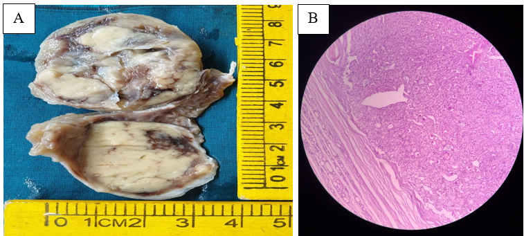
a): Gross -single grey white capsulated nodule; b): Microscopic examination showed microfollicles with fibrous capsule

Table 4
Age, Sex and histological categories of all thyroid lesions
Analysis of sex showed female predominance of 87.5% similar to Fatima et al at 89.2% and 90% in Ramesh et al 9 and Jagadale et al.10 Non-neoplastic lesions in present study were 72% and neoplastic were 29% which correlated with Jagadale et al 71.4% of non neoplastic lesions. However, Ramesh V.C. et al9 found 47.5% and Fatima et al8 found 83.3% of non neoplastic lesions respectively (Table 5).
Table 5
Comparison of non-neoplastic lesions and neoplastic lesions of thyroid
| Thyroid lesions | Ramesh VL (n=120) | Jagadale K (n=70) | Fatima et al (n=120) | Present study |
| Non neoplastic | 47.5% | 71.4% | 83.33% | 72% |
| Neoplastic | 52.5% | 28.6% | 16.67% | 28% |
Comparing the non-neoplastic lesions colloid goiter (32.6%) and multinodular goiter (27.8%), Hashimoto’s thyroiditis (8%) were the common lesions.
Our present study correlated well with Jagadale K et al10 with colloid goiter 21.4%, MNG 28.6% and Hashimotos thyroiditis 8.57%. Comparision with other studies for non neoplastic lesions is shown in Table 6.
Table 6
Comparison of non-neoplastic lesions of thyroid
Analysis of neoplastic lesion showed follicular adenoma 14 cases (13.4%), followed by papillary thyroid carcinoma 13(12.5%), Follicular carcinoma 2cases (1.9%), WDFT-UMP 1case (0.9%), NIFTP 1case (0.9%) Analysis of neoplastic lesions show predominance in females (Figure 3). Comparison with other studies is shown in the Table 7.
Table 7
Comparison of neoplastic lesions of thyroid
| Neoplastic lesion | Ramesh V L (n=120) 2014 | Jagadale K (n=70) 2016 | Fatima et al (n=120) 2016 | Present study (n=104) 2019 |
| Follicular adenoma | 36% | 7.2% | 12.5% | 13.4% |
| Papillary carcinoma | 15% | 8.7% | 2.5% | 12.5% |
Out of 29 cases of Multinodular goiter in our study, 2 cases showed incidental papillary carcinoma. Similarly, Jain et al found 3 cases of papillary microcarcinoma out of 35 cases operated for multinodular goiter.11, 12 Kumar et al found malignant foci in 21 cases (8.1%) among 258 clinically diagnosed nodular goiter in which papillary carcinoma was the most common type of malignancy.12 Hence thorough screening of all thyroidectomy specimens to rule out occult carcinoma as the risk of carcinoma in MNG is significant.
Conclusion
The present study was concluded with the following observations
Neoplastic and non neoplastic lesions were common in females 87.5%.
Common age group affected 2nd to 3rd decade.
Commonest non neoplastic lesion was Colloid goiter (32.6%) followed by multinodular goiter (27.8%).
Commonest benign neoplasm was follicular adenoma (13.4%).
Commonest malignant neoplasm was papillary carcinoma(12.5%).
The present study highlights the importance of histopathological typing of thyroid lesions for their better management.
All thyroidectomy specimens should be throughly grossed to rule out occult malignancy as the risk of carcinoma in Multinodular goiter is significant.


