Introduction
Lung carcinoma is the most common and lethal cancer in men3, 2, 1 attributed to tobacco consumption. Non-small cell carcinomas (NSCLC) are more common and aggressive than small cell lung carcinomas. Most commonly lung cancers presentsat an advanced stage.4 Lung carcinomas are histologically and molecularly heterogeneous, hence rapid and meticulous work-up is often mandated. Immunohistochemistry and metabolic studies like 18F-FDG PET CT scan plays an important role in the initial assessment and diagnostic confirmation. The 2015 World Health Organization (WHO) classification is a better modified version, based on molecular marker study and genetic alterations in lung cancer. Molecular markers in case of lung carcinoma are EGFR mutation, KRAS mutation, ROS 1 gene rearrangement and Her2. The current guidelines recommend EGFR, ALK and ROS 1 testing. Targeted therapy can be initiated in cases of positive EGFR, ROS 1 or ALK genetic rearrangements.
Here in presenting a case report of TTF 1 and Napsin A negative lung adenocarcinoma with elevated serum CA19-9, imposing the necessity of pathologico-clinico-radiological correlation, especially in advanced cancers.
Case Report
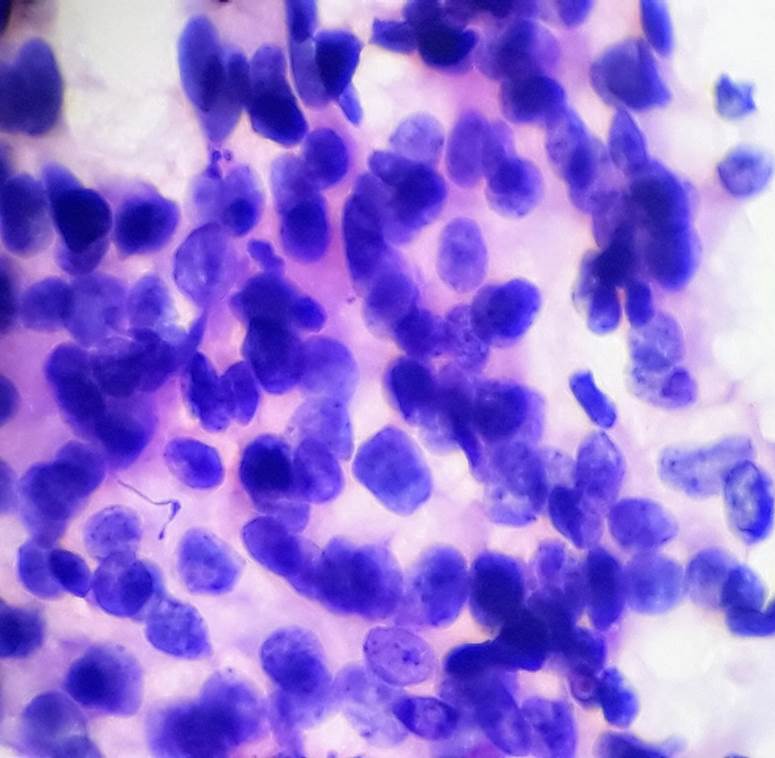
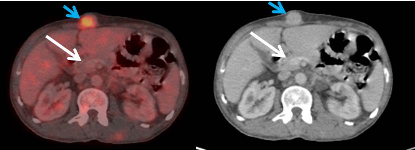
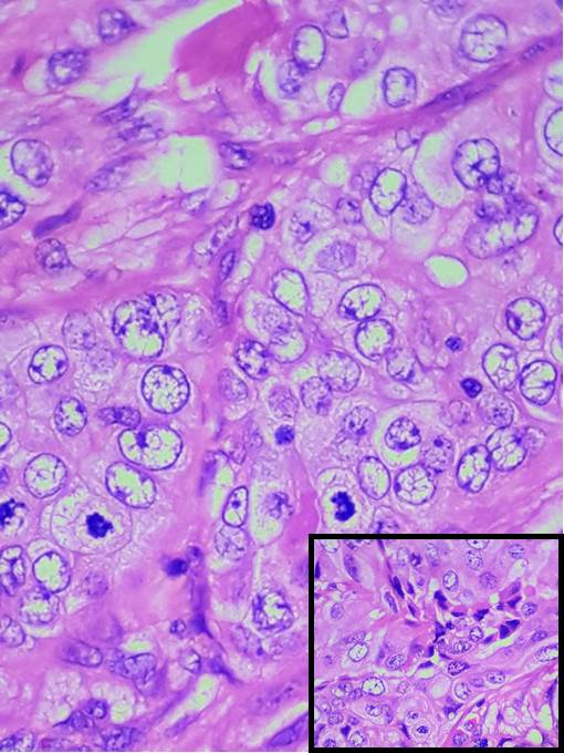
A 56 year old male presented with multiple subcutaneous nodules of varying sizes all over the body. (Figure 1). Clinical examination revealed cachexic, anicteric and anaemic individual. Nodule size varied from 4 x 3 cms to 1 x 1 cm. Nodules were mobile, firm to hard in consistency and non-tender. Systemic examination revealed no organomegaly and no neurological deficit. Haematological investigaton revealed severe anaemia. Biochemical parameters were within normal limits. FNAC of subcutaneous nodules was suggesting metastatic adenocarcinoma deposits. (Figure 2). Incision biopsy of subcutaneous nodules was advised along with 18F-FDG PET CT scan. Incision biopsy of subcutaneous nodules was consistent with metastatic adenocarcinoma deposits. (Figure 4, Figure 3). 18F-FDG PET CT revealed metabolically active right pleural based mass lesion (8.1 x 6.1 x 6.3 cm) in posterior aspect of right lower zone lung. (Figure 6, Figure 5). Multiple metabolically active pleural based nodular mass in right hemithorax, mediastinal lymphadenopathy and muscular deposits were also revealed.
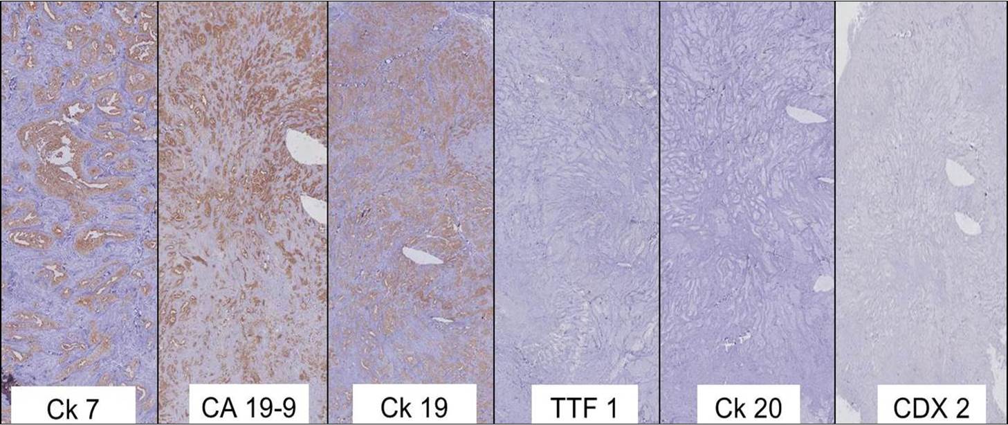
Immunohistochemical study showed positivity for C k7, CA 19-9 and Ck19 and immunonegative for TTF 1, Napsin A, p40, Ck20, CDX-2, GATA-3 and Calretinin. (Figure 7). Possibility of adenocarcinoma of pancreatic origin was suggested on IHC of subcutaneous nodule.
18F-FDG PET CT scan and IHC were diverging/ dichotomous in the localisation of primary tumour. The dilemma existed between TTF 1 and Napsin A negative metastatic lung adenocarcinoma and CA 19-9 positive metastatic pancreatic adenocarcinoma. After review of all reports further tests were advised to aid in localisation of primary tumour. Serum CA 19-9 and EGFR mutation analysis by IHC, ROS 1 gene rearrangement by IHC and ALK by IHC were advised. Serum CA 19-9 was 4380 U/ml. EGFR mutation analysis by IHC showed Wild Type, ROS 1 gene rearrangement by IHC was positive and ALK (D5F3) by IHC was negative. Considering all the metabolic imaging, histopathological and IHC panel confirmation, final diagnosis was considered by exclusion and inclusion as lung adenocarcinoma and ROS 1 targeted therapy was started. Radiation therapy was not contemplated as the disease was already metastatic at the presentation and no pacemaker lesion that required palliation could be identified at that point of time. Treatment with Crizotinib was initiated.
Figure 3
Microphotograph of histopathology from subcutaneous nodule showing metastatic adenocarcinoma deposits
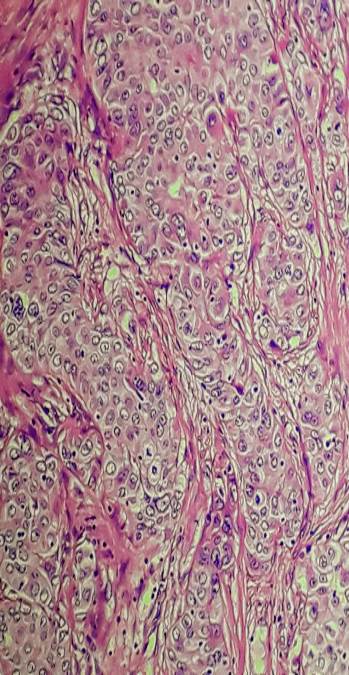
Figure 4
Microphotograph of histopathology from subcutaneous nodule showing metastatic adenocarcinoma deposits
Discussion
Lung cancer is the most common cancer in men and third most common cancer in women.1 It is one of the common lethal diseases afflicting the global population.2 Cigarrete and other forms of tobacco consumption attributes to most of the cases of lung cancer. Lung carcinomas were classified broadly into small cell carcinoma and non-small cell lung carcinoma (NSCLC) till WHO 2004 classification. Non-small cell carcinomas are more common and aggressive as compared to small cell carcinoma of lung.5, 3 Prior to WHO 2004 classification, NSCLCs tumors were not classified further in small tissue samples because of therapeutic options paucity. WHO 2004 classified NSCLCs into squamous cell carcinoma (SqCC), adenocarcinoma (ADC) and others. WHO 2015 further classified NSCLCs. It emphasised the use of immunohistochemistry and genetic studies. Integration of molecular testing helped to personalize treatment strategies, especially for advanced lung cancer patients.6
Platinum based chemotherapy is the standard of care for NSCLC with Permetrexed being used in adenocarcinoma. According to WHO current guidelines testing for EGFR, ALK and ROS 1 is essential for all lung adenocarcinomas.
Most lung cancers present at an advanced stage with aggressiveness and metastatic potential.4 Cytology and histopathology plays an important role in the initial assessment of these tumours especially those at advanced stage with metastatic deposits.7 After the initial assessment, localization of primary tumour is mandatory for initiation and planning therapy. Immunohistochemistry, radiology and metabolic imaging plays crucial role in diagnosis and localization of primary tumours.
The emerging immunohistochemical markers preferred as classifier of NSCLC include TTF 1 and Napsin A (for ADC) and p40, p63 and CK5/CK6 (for SqCC). Sensitivity and specificity of TTF 1 and Napsin A ranges from 80%-90% in cases of adenocarcinoma lung. Sensitivity of Napsin A is higher as compared to TTF 1.8 The positivity of both TTF 1 and Napsin A decreases with increasing tumour grade.7 Similar decrease in positivity in metastatic lung ADC were seen in study done by Gurda et al,7 where TTF1 expressed the sensitivity and specificity of 84.5% and 96.4% in cases of primary lung ADCs, and 86.9% and 87.5% in metastatic lung ADCs. Napsin A expressed sensitivity and specificity of 92.0% and 100% in the primary lung ADCs, and 67.8% and 100% in cases of metastatic lung ADCs.
NSCLC should be tested for epidermal growth factor receptor (EGFR) mutations, anaplastic lymphoma kinase (ALK) and proto-oncogene tyrosine-protein kinase ROS 1 (ROS 1) gene rearrangement. The presence of these mutations predicts the responsiveness to tyrosine kinase inhibitors (TKIs) and Crizotinib.
Carbohydrate antigen 19-9 (CA 19-9) is isolated from a human colorectal cancer cell line.9, 7 Although applied in the diagnosis of pancreatico- biliary adenocarcinoma, it can be positive in advanced lung adenocarcinoma (ALADC) also.10 CA 19-9 is extremely elevated in advanced lung adenocarcinoma. This spurious and deceptive high value of CA 19-9 can cause diagnostic dilemma in cases of non-pancreatic adenocarcinoma. Similar elevation of CA 19-9 was seen in the present case of advanced lung adenocarcinoma.
Metabolic imaging combined with radiological imaging is rapidly progressing modality of choice, especially in oncology set-ups. The metabolic uptake of the glucose analog 18F-FDG is studied in Positron emission tomography (PET) scan. Combination of PET with computerised tomography (CT) scan increases the sensitivity and specificity to 85% - 95%.12, 11 Being economical tool in the evaluation of lung cancer, 18F-FDG PET stands ahead in diagnosing, staging, and evaluating response to treatment.10
The present case is lung carcinoma with multiple metastatic deposits. Initial assessment of metatstatic adenocarcinoma by cytology and histopathology was supported by IHC which showed negativity for TTF 1 and Napsin A and positivity for CA 19-9 and Ck 19, suggesting adenocarcinoma of pancreatico-biliary origin. 18F-FDG PET CT scan revealed metabolically active right pleural based mass lesion (8.1 x 6.1 x 6.3 cm) in posterior aspect of right lower zone lung with multiple pleural nodular mass in right hemithorax, mediastinal lymphadenopathy and muscular deposits. Immunohistochemistry of subcutaneous nodule was negative for TTF 1 and Napsin A and immunoreactive for CA 19-9 with markedly elevated serum CA 19-9. Working as a team of pathologist, oncologist and radiologist we could diagnose the case of TTF 1 and Napsin A negative lung adenocarcinoma with elevated CA 19-9.
Conclusion
Advanced tumours requests vigilant and meticulous investigations to be carried out. CA 19-9 elevation can be seen in non-pancreatic ADC,10 such as lung ADC in the present case. Its elevation is a bad prognostic marker. Napsin A and TTF 1 positivity decreases with increasing tumor grade of Lung ADC. A physician/ pathologist should not be biased, especially in advanced stage cancers. Clinico-pathologico-radiological correlation is mandatory. Multidisciplinary approach and team work of pathologist, oncologist and radiologist helps in concluding diagnostic dilemmas, especially in case of advanced carcinomas.


