- Visibility 246 Views
- Downloads 30 Downloads
- Permissions
- DOI 10.18231/j.ijpo.2019.124
-
CrossMark

Abstract
Introduction: The lacrimal gland is non-capsulated, multilobulated secretory gland situated in the lacrimal gland fossa. Lesion and tumors of lacrimal gland occupy almost 18% of all orbital tumors. Pleomorphic adenoma of lacrimal gland also known as mixed tumour of lacrimal gland it is the most common tumor as well as most common benign tumor of lacrimal gland and very critical to differentiate between adenocystic and pleomorphic adenoma of lacrimal gland because of different prognosis.
Aims and Objectives: To study the prevalence of pleomorphic adenoma in to the lacrimal gland tumors and to study the histopathological pattern of pleomorphic adenoma in lacrimal gland, its distribution into the age, sex and its outcome in to different condition.
Materials and Methods: A retrospective study of 38 cases of pleomorphic adenoma in lacrimal gland during period of 10 years were studied at tertiary care institute. A detailed history of patients were taken with special attention to the age, sex, macroscopic (gross examination) and microscopic features of tissue. Microscopic examination is done under standard H & E staining procedure. Microscopic examination is focused on specifically tumour cells size, shape, nucleus characteristics and cytoplasm and arrangement of cells.
Result: Total 57 cases of lacrimal gland tumor were studied among them 38 cases (66.66%) of pleomorphic adenoma were diagnosed. Among 38 cases of pleomorphic adenoma 26 cases (68.4%) were in Female and 12 cases (31.6%) were in male. Among them most number of cases are seen in the age group of 21-40 years almost 18 cases (47.3%), and second common frequency in to the 41-60 years age group contain 13 cases (34.2%). When studied according to histological features, then Myxoid differentiation are seen in 33 (94%)cases, Squamous metaplasia are seen in 18 cases(47%), Chondroid differentiation are seen in 16 cases(42%), Cystic degeneration are seen in 12 cases (33%), Calcification are seen in 3 cases (8%), Cellular atypia are seen in 2 cases(6%), Osseous metaplasia is seen in 1 case (2.6%), Hy aline changes are seen S in 17 cases (45%), Predominantly cellular appearance in 21 cases (55.2%), Predominantly stromal appearance in 1 cases (2.6%), Mixed (cellular + stromal) 3 cases (7.9%).
Conclusion: Pleomorphic adenoma is most common benign tumour of lacrimal gland tumour and its incidence are significantly higher into the female (68.4%) and most common presenting age group is 21-40 (47.3%) and if bar is raised to 21-60 years then almost 80% cases fall in this category so most commonly seen in middle age group.
Introduction
The lacrimal gland is non-capsulated, multilobulated secretory gland situated in the lacrimal gland fossa. Lesion and tumors of lacrimal gland occupy almost 18% of all orbital tumor.[1] Lacrimal gland tumor are relatively rare so most publish article describe single case or small series, But in our study we studied almost 38 cases in duration of more than 10 years. Structure of lacrimal gland is similar to salivary gland so for the classification of the lacrimal gland tumor is done according to the WHO (world health organisation) classification of salivary gland tumors.[2]
Who classification of salivary gland tumors
Malignant epithelial tumors
Acinic cell carcinoma
Mucoepidermoid carcinoma
Adenoid cystic carcinoma
Polymorphous low grade adenocarcinoma
Epithelial-myoepithelial carcinoma
Clear cell carcinoma, not otherwise specific
Basal cell adenocarcinoma
Sebaceous carcinoma
Sebaceous lymphadenocarcinoma
Cystadenocarcinoma
Low grade cribriform cystadenocarcinoma
Mucinous adenocarcinoma
Oncocytic carcinoma
Salivary duct carcinoma
Adenocarcinoma, not otherwise specified
Myoepithelial carcinoma
Carcinoma ex pleomorphic adenoma
Carcinosarcoma
Metastasizing pleomorphic adenoma
Squamous cell carcinoma
Small cell carcinoma
Large cell carcinoma
Lymphoepithelial carcinoma
Sialoblastoma
Benign epithelial tumors
Pleomorphic adenoma
Myoepithelioma
Basal cell adenoma
Warthin”s tumor
Basal cell adenoma
Oncocytoma
Canalicular adenoma
Sebaceous adenoma
Ductal papilloma
Inverted ductal papilloma
Intraductal papilloma
Sialadenoma papilliferum
Cyst adenoma
Lymph adenoma
(a) Sebaceous (b) Non-sebaceous
Secondary tumors
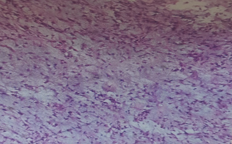
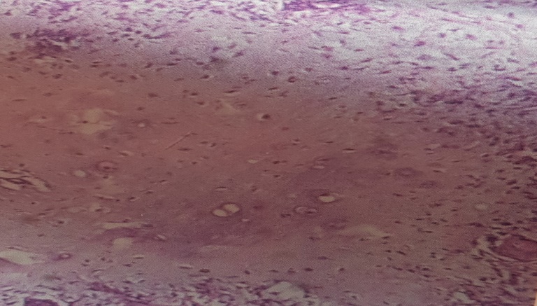
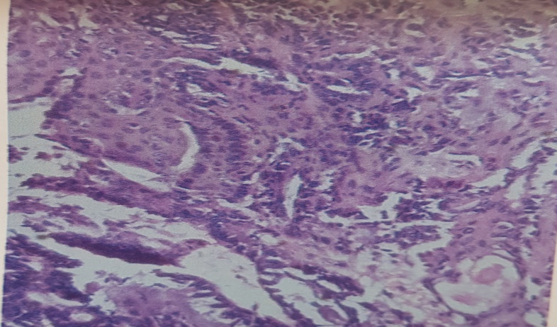
Pleomorphic adenoma of lacrimal gland is a well define benign tumour characterised by a mixed appearance of proliferated epithelial ducts and myxoid, mucoid or cartilaginous area containing myoepithelial cells. Tumour arise from progenitor ductal cell capable of both epithelial and myoepithelial differentiation.[1] Pleomorphic adenomas the most common primary epithelial tumor of lacrimal gland and arise most commonly from orbital lobe (90%), and occasionally from palpebral lobe and rarely from accessory lacrimal gland.[3],[4],[5] Most patient present with slowly progressive, painless mass at the supratemporal orbit. Grossly pleomorphic adenoma are pseudo capsulated, smooth surfaced tumours and cut surface shows alternating mucinous and fibrous areas with occasional cystic structure. A layer of compressed connective tissue surrounds the tumor and focal small tumor projections known as “BOSSELATIONS”. Microscopically characteristic features are the presence of epithelium – lined duct like structure and outer layer of darker myoepithelial cells. Mucoid, basophilic material accumulate among the myoepithelial cells and between the epithelial ducts like structure. In some cases foci of chondroid like material and even calcification are observed. The epithelial cell may form solid cords and even show squamous differentiations and cyst formations. The microscopic diagnosis of Pleomorphic adenoma is easy but sometimes confuse with myoepithelial adenoma and oncocytic adenomas. Pleomorphic Adenoma are DNA diploid tumor with low expression of the p53 gene.[6] P21 Expression is apparently related to progression of the pleomorphic adenoma of the lacrimal gland.one study showed recurrent chromosomal abnormalities involving chromosome 3, 8, 9, and 12 similar to those found in salivary gland tumours.[7],[8] Sensitivity of preoperative diagnosis is approximately 90%. The overall 15 year recurrence rate is less than 3% if the tumor removed at the initial surgery. Despite the presence of recurrence the survival rate at 15 years is almost 100%.
Materials and Methods
A retrospective study of 38 cases of pleomorphic adenoma of lacrimal gland tumours were studied over a period of 10 years at tertiary care institute. A detailed clinical history was obtained with specific focus on age, sex and histopathological examination include macroscopic examination and microscopic examination with special stains like PAS (periodic acid Schiff ’s) stain and immunohistochemistry where it required. For macroscopic examination specimen were fixed in 10% buffered formalin and then processed, sectioned and stained with H & E (haematoxylin and eosin stain) stain. For Microscopic examination H & E stained sections were studied from different areas. Sections were studied for diagnosis of tumor, others histological features and differention and for individual tumor cells type which includes size, shape, characters of nucleus, nucleolus.
Result
Total 57 cases of lacrimal gland tumor were studied among them pleomorphic adenoma consist of 38 cases (66.66%) cases. Other tumor were diagnosed are adenocystic carcinoma, mucoepidermoid carcinoma, squamous cell carcinoma but in this study our focus on pleomorphic adenoma.so prevalence of pleomorphic adenoma is 66.66%. Present study consists of 38 cases of pleomorphic adenoma of lacrimal gland. When studied according to the age group, then 0-20 years groups consist of 2 cases (5.3%), 21-40 years age group consist of 18 cases (47.3%), 41-60 years age groups consist of 13 cases (34.2%) cases, and > 60 years age group consist of 5 cases (13.2%). When 38 cases studied according to sex then out of 38 cases, 26 cases (68.4%) were in female and 12 cases (31.6%) were in males. On gross examination Pleomorphic adenoma typically have a pseudo capsule surrounding the mass lesion. Histologically, pleomorphic adenoma shows varying proportions of epithelial and mesenchymal components. The epithelial cells form characteristic double layer ducts, acini, strands, irregular tubules and surrounding myoepithelial cells with background of loose myxoid tissue containing chondroid and rarely bone formation. When divided accordingly histological features then Myxoid differentiation in 33(94%)cases, Squamous metaplasia in 18 cases (47%), Chondroid differentiation in 16 cases(42%), Cystic degeneration in 12 cases (33%), Calcification in 3 cases (8%), Cellular atypia in 2 cases(6%), Osseous metaplasia in 1 cases (2.6%), Hyline changes in 17 cases (45%), Predominantly cellular appearance in 21 cases (55.2%), Predominantly stromal appearance in 1 cases (2.6%), Mixed (cellular + stromal) (3) 3 cases (7.9%).
| Age group (years) | Number | Percentage (%) |
| 0-20 | 2 | 5.3% |
| 21-40 | 18 | 47.3% |
| 41-60 | 13 | 34.2% |
| >60 | 5 | 13.2% |
| Total | 38 | 100% |
| Age group (years) | No of cases in Male | No of cases in Female | Total no case |
| 0-20 | 0 | 02 | 02 |
| 21-40 | 6 | 12 | 18 |
| 41-60 | 4 | 09 | 13 |
| >60 | 2 | 03 | 05 |
| Total | 12 (31.6%) | 26 (68.4%) | 38 |
| Myxoid differentiation | 33 | 94% |
| Squamous metaplasia | 18 | 47% |
| Chondroid differentiation | 16 | 42% |
| Cystic degeneration | 12 | 33% |
| Calcification | 3 | 8% |
| Cellular atypia | 2 | 6% |
| Osseous metaplasia | 1 | 2.63% |
| Hyline changes | 17 | 45% |
| Predominantly cellular | 21 | 55.26% |
| Predominantly stromal | 1 | 2.63% |
| Mixed (cellular + stromal) | 3 | 7.9% |

Discussion
A study of 38 cases of pleomorphic adenoma of lacrimal gland over 10 years was done. when cases are divided according to age and sex then results are summarised in [Table 2]. So among total 38 cases the maximum cases in age group is 21-40 years age group ([Table 1]) contain 18 cases (47.3%) as some study[9],[10] suggest that pleomorphic adenoma is most common in middle age, second age group which contain most cases is 41-60 years age group contain 13 cases (34.2%), and after that > 60 years age group contain 5 cases (13.2%), and followed by 0-20 years contain 2 cases (5.3%). So pleomorphic adenoma is least common in early age in childhood in our study. Secondly when focus in sex distribution of tumor in specific age group as well as in total cases result is out of 38 cases total 26 cases (68.4%) are seen in female and 12 cases (31.6%) are seen in male. And when divided into specific age group and then incidence noted according to sex then also individual age group shows clear cut female predominance [Table 2]. Now on when focused on various histopathological features on microscopy study results are following. On microscopy 33 cases (94%) cases shows myxoid differentiation, 16 cases(42%) shows chondroid differentiation, 17 cases (45%) shows hyaline changes, 18 cases(47%) shows squamous metaplasia, [Figure 4] 3 cases (8%) shows calcification, 2 cases (6%) shows cellular atypia, 12 cases (33%) shows cystic degenerative changes, 1 case(2.6%) shows osseous metaplasia [Table 3]. Other histological changes are also detected but they are not in notifiable amount. Among 38 cases 21 cases are predominantly cellular component are seen, when in 1 cases predominantly stromal component are seen, and in 3 cases mixed pattern are seen. So on above result the most common histological change is noticed is the myxoid differentiation (94%), second most common squamous metaplasia in 47% cases, followed by hyaline change (45%), and chondroid differentiation (42%). So for the summary the pleomorphic adenoma is relatively common in female and common presenting age group is middle age and most common histological feature present is the myxoid differentiation.
Conclusion
A retrospective study of 38 cases of pleomorphic adenoma of lacrimal gland shows pleomorphic adenoma is the most common benign tumor of the lacrimal gland as well as most common tumor of the lacrimal gland. The peak age for pleomorphic adenoma is the middle age mostly 21-40 year age groups contain 47.3% cases, if bar is raised and group is made 21-60 years middle age groups then more than 80% or exactly 81.5% cases fall in that group and extreme of age means in childhood and old age this tumour is less prevalent but incidence are there. And after that focus on gender we found that out of 38 cases 26 cases are seen in female means 68.4% cases are seen in female show it indicate slight female preponderance not even in total cases but in individual age group case also shows female preponderances so pleomorphic adenoma is slight more prevalent in to the Female. On comment in histological features on microscopy shows most common histological features noted was myxoid differentiation which is seen in 94% cases, followed by the squamous metaplasia in 47% cases, other histological features are in less significant amount.
Source of funding
None.
Conflict of interest
None.
References
- Iwamoto T, Jakobiec FA. A comparative ultra structural study of the normal lacrimal gland and its epithelial tumors. Hum Pathol. 1982;13:236-262. [Google Scholar]
- JFL. Tumors of the Eye and Ocular Adnexa. Vol 55th ed. Washigton, DC American Registry of pathology and Armed forces institute of pathology 2006. . . [Google Scholar]
- Parks SL, Glover AT. Benign mixed tumor arising in the palpebral lobe of the lacrimal gland. Ophthalmol. 1990;97:526-562. [Google Scholar]
- Vangveeravong S, Katz SE, Rootman J, White V. Tumors arising in the palpebral lobe of the lacrimal gland. Ophthalmol. 1996;103:1606-1612. [Google Scholar]
- Tong JT, Flanagan JC, Eagle RC, Mazzoli RA. Benign mixed tumor arising from an accessory lacrimal gland of wolfring. Opthal Plast Reconstr Surg. 1995;11:136-149. [Google Scholar]
- Zho P, Sun X. Quantitative analyses of DNA content and P53 gene product expression from epithelial lacrimal gland tumors. Zhonghua Yan Ke Za Zhi. 1996;32:424-428. [Google Scholar]
- Jin Y, Mertens F, Limon J, Mandahl N, Wennerberg J, Dictor M. Characteristic karyotypic features in lacrimal and salivary gland carcinomas. Br J Cancer . 1994;70:42-47. [Google Scholar]
- Hrynchak M, White V, Berean K, Horseman D. Cytogenetic finding in Seven Lacrimal Gland Neoplasm. Cancer Genet Cytogenet. 1994;75:133-138. [Google Scholar]
- Rl -, Gamel JW, . JF. Epithelial tumors of the lacrimal gland: Analysis of 265 cases. Ocular and Adnexal tumor. 1978. [Google Scholar]
- Rose ER, Wright JE. Pleomorphic adenoma of the lacrimal gland. Br J Ophthalmol. 1992;76:395-395. [Google Scholar]