Introduction
Soft tissue encompasses the supportive connective tissue of various organs and the other nonepithelial, extra skeletal structures excluding the viscera, lymphoreticular system and coverings of the brain (meninges). It is represented by adipose tissue, fibrous connective tissue, peripheral nervous system, skeletal muscle and blood/lymph vessels.1
Most of the soft tissues originate embryologically from the mesodermal layer.
Benign mesenchymal tumours are commoner than the malignant ones and outnumber them by a ratio of almost 100:1.2 But the fact that most of the benign lesions like lipomas and haemangiomas, are not investigated and biopsied, makes it difficult to apply data from many hospital studies for the general population.3
Soft tissue sarcomas comprise less than 1% of all the malignant tumours. Due to the rarity of these tumours, there is a paucity of the literature regarding the soft tissue sarcomas especially among the developing countries.4
The pathogenesis of most of the soft tissue tumours is still unknown.5 Some of the known causes are irradiations, immune deficiency, genetic factors, viral infections and environmental factors.
Even though soft tissue tumours may appear at any age, there is a relation between the patient’s age, gender, location and type of tumour.6,7
Both benign and malignant soft tissue tumours most commonly present as slowly growing painless mass. A sudden and rapid enlargement of a soft tissue mass must arise a suspicion of a malignant lesion.1
Soft tissue tumours have been seen to arise almost anywhere in the body, but the most frequent locations include extremities, head and neck region, trunk and abdominal cavity.8
The major indications of biopsy in case of soft tissue lesions are a mass arising with or without a history of trauma and a mass present beyond 6 weeks after local trauma. An open biopsy is considered as the gold standard in diagnosing soft tissue neoplasms.9
A microscopic examination of the haematoxylin & eosin stained sections remains the gold standard in diagnosing soft tissue tumours.
The diagnostic accuracy can be increased by application of special techniques like electron microscopy, conventional special stains, cytogenetics, immunohistochemistry and molecular methods.10 Availability now of an updated histogenetic classification (WHO classification) and a standard nomenclature offers a good clinicopathological correlation. The aim and objectives of this study are:
Materials and Methods
This was a prospective study of 100 cases conducted at the Department of Pathology at Dr. D. Y. Patil Medical College, Hospital and Research Centre, Pune, Maharashtra. Institute Ethics Committee Clearance (IECC) was obtained before the start of the study. Detailed clinical history, important clinical examination findings, radiological findings were obtained and noted on a structured proforma.
All specimens were received in 10% buffered formalin and were processed through the standard paraffin embedding technique. Sections of 3-5 micrometre thickness were cut and stained with Haematoxylin and Eosin stain. Special stains like PAS, Reticulin and IHC were done wherever necessary. The sections were examined under the microscope and classified as per the 2013 WHO classification.
Results
Total 100 cases of soft tissue tumours were included in our study.
Table 1
Incidence of benign, intermediate and malignant soft tissue tumours
Table 2
| Site | Benign | Intermediate | Malignant | Total |
| Extremities | 29 | 0 | 6 | 35 |
| Head and neck | 32 | 0 | 0 | 32 |
| Back and shoulder | 18 | 0 | 0 | 18 |
| Trunk and abdomen | 9 | 2 | 2 | 13 |
| Others | 2 | 0 | 0 | 2 |
| Total | 90 | 2 | 8 | 100 |
Site wise distribution of benign, intermediate and malignant soft tissue tumours
Benign soft tissue tumours formed 90% of all soft tissue tumours, while 2% were intermediate and malignant soft tissue tumours comprised an 8% among all the soft tissue tumours. The benign to malignant ratio was 11.3 (Figure 1).
The soft tissue neoplasms overall had a marginal male predominance with a male to female ratio of 1.3:1. In the benign lesions, it was 1.3:1 while among the malignant tumours it was 1:1.7, that is, sarcomas showed a female predominance. (Figure 1)
Soft tissue tumours were seen most commonly in the fifth decade (23%) followed by the third decade (19%) (Figure 2).
The benign soft tissue tumours had a peak incidence in the fifth decade. While the malignant soft tissue tumours were seen to occur in the age group above 60 years of age.
On histopathological examination of the soft tissue tumours, most common histological type was adipocytic tumours, which constituted 55% of all the tumours, followed by nerve sheath tumours (18%) and vascular tumours (14%) (Table 1).
Lipoma (5 4.4 %) was the most common benign tumour of soft tissue (Table 1).
Amongst the intermediate grade of soft tissue tumours, one was a case of atypical lipomatous tumour, while the other case was a fibrosarcomatous variant of DFSP.
The most commonly encountered malignant soft tissue tumours were liposarcomas and undifferentiated pleomorphic sarcomas (25% each).
Benign soft tissue tumours had a predilection for the head and neck region, followed by extremities (Table 2).
Malignant soft tissue neoplasms had a predilection for the lower extremities (Table 2).
Figure 1
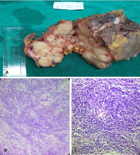
DFSP, fibrosarcomatous variant: (Gross, H&E, x100 & x400) A): (Gross)- showing well-circumscribed, large, whitish, lobular soft to firm mass; B): Photomicrograph showing tumour tissue arranged in a predominantly storiform or herringbone pattern. (H&E, x100); C): Photomicrograph showing spindled tumour cells containing plump or elongated wavy nuclei with pleomorphism and many hyperchromatic nuclei. Few atypical mitotic figures noted. (H&E, x400).

Figure 2
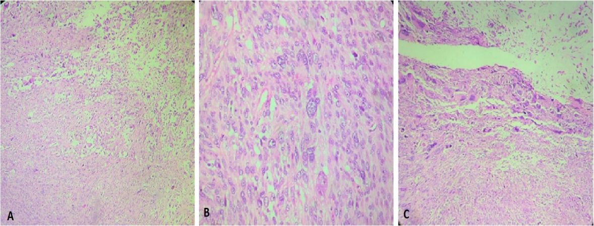
Pleomorphic liposarcoma: (H&E, x100 & x400); A): Photomicrograph showing spindled cells and multinucleate tumour giant cells arranged in short fascicles. (H&E, x100); B): Showing pleomorphic hyperchromatic nuclei with vesicular chromatin and prominent nucleoli with few atypical mitotic figures. (H&E, x400); C): Showing extremely large lipoblasts with clear or vacuolated cytoplasm and central nucleus. (H&E, x400).

Figure 3
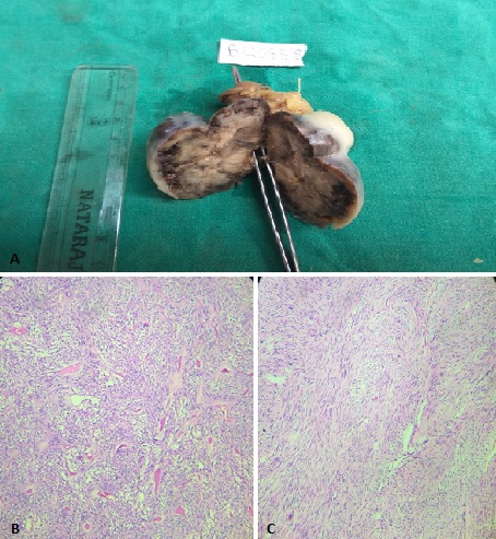
GIST: (Gross, H&E, x100 & x400); A): (Gross)- showing tumour mass which is greyish white with few dark areas, solid and firm in consistency; B): Photomicrograph showing spindle shaped cells with epithelioid cells at places and occasional skeinoid fibres and hyalinized blood vessels. (H&E, x100); C): Photomicrograph showing tumour tissue having classical spindle cell morphology and arranged in fascicles and organoid pattern. (H&E, x100).

Figure 4
Undifferentiated pleomorphic sarcoma: (H&E, x100 & x400); A): Photomicrograph showing tumour tissue arranged in fascicles with prominent rhabdoid differentiation. (H&E, x100); B): Showing tumour cells having plump, spindeloid and round cells with large hyperchromatic pleomorphic nuclei with nucleoli & few atypical mitotic figures. (H&E, x400); C): Showing multinucleate tumour giant cells with bizarre hyperchromatic nuclei. (H&E, x100).

Discussion
Soft tissue tumours, both benign and malignant, are those fascinating group of lesions which arise from the supportive soft tissue of the body and have a very broad spectrum not only of the histological entities, but also of their behaviour and histomorphologic features with close histopathologic similarities between certain tumours and having only subtle differences which are detectable on a detailed microscopic examination, thus posing a diagnostic challenge to the histopathologist.
Soft tissues comprise the nonepithelial extra skeletal tissues of our body, which does not include the reticuloendothelial system, glia and supporting tissues of various parenchymal organs. They are represented by adipose tissue, fibrous tissue and voluntary muscles along with the vessels supplying these tissues.
Table 3
Distribution of soft tissue tumours in various studies
In the present study, we analysed 100 cases of soft tissue tumours out of which 90% were benign, 2% were intermediate and 8% were malignant, showing that benign tumours were most common followed by malignant tumours. Intermediate category of tumours were the least commonly encountered tumours in the study. These proportions were similar to those reported by Jain P et al,5 Umarani MK et al12 and Swagata D et al,13 Narayanan NO et al1 studied 291 cases of soft tissue tumours and reported 93.8% benign, 3.4% intermediate and 2.8% malignant tumours. A retrospective study by Peterson et al11 showed 49% malignant, 35% benign, 11.4% intermediate and 4.6% of tumours with uncertain potential.
In all these studies, benign tumours have been seen to predominate the malignant ones, although the relative frequency of benign to malignant soft tissue tumours till date has been difficult to estimate. This could be because the benign soft tissue tumours do not cause many complaints to the patients and hence, they do not usually report to the clinician or surgeon and these benign tumours are thus not surgically excised.
Soft tissue tumours have a varied age distribution and all ages are at risk for developing these lesions. The majority of cases in the present study were seen in the fifth decade (23%) followed by the third decade (19%). We reported haemangiomas to be the most common tumour of childhood. We found that the sarcomas were more common in the elderly and had a peak incidence in the 6th decade and beyond (50%). Similar findings were reported by Narayanan NO et al,1 Baste BD et al14 and Janaki M et al.15
In our study, we had 56 males and 44 females with a male to female ratio of 1.3:1 which is same as the ratio of Swagata D et al13 and Jain P et al.5 Our findings are also similar to the study done by Umarani MK et al12 who found the male to female ratio to be 1.1:1. A male predominance was noted in almost all the soft tissue tumours among the various studies available. The male to female ratio in benign soft tissue tumours in the present study was 1.3:1; while among the malignant tumours it was 1:1.7, that is the malignant tumours showed a female preponderance. The reason for this could be that females generally tend to ignore their health issues and hence present to the clinician at a late stage when there has been a rapid and massive increase of the lesion which poses many difficulties in their day to day activities.
Among the benign soft tissue tumours, adipose tissue tumours (57.8%) formed the largest group followed by nerve sheath tumours (18.9%) and vascular tumours (15.6%). Similar findings were reported by Umarani MK et al12 and Baste BD et al.14 There has been noted a highly significant association between the type and category of soft tissue tumours. Gastrointestinal stromal tumours (GIST) and tumours of uncertain potential (1.1% each) constituted the least commonly encountered tumours in our study. Most of the benign tumours in our study were located in the head and neck region (35.6%), followed by the extremities (32.2%). Various studies available in literature like Narayanan NO et al,1 Jain P et al,5 Baste BD et al, etc. found extremities to be more commonly involved than the head and neck region. The reason for higher proportion of benign tumours in head and neck region could be that since our study was conducted in an urban area, patients reported more frequently to us with head and neck tumours due to cosmetic reasons.
Liposarcomas and undifferentiated pleomorphic sarcomas (25% each) were the most common malignant soft tissue tumours in our study. Our findings were in accordance with Baste BD et al14 and Samartha V et al.16 Majority of the soft tissue sarcomas were located in the extremities (75%), especially the lower extremities (62.5%), followed by the trunk and abdomen (25%). Similar findings were reported by Samartha V et al,16 Jain P et al5 and Narayanan NO et al.1 However, the study conducted by Mandong et al17 reported extremities (45.7%) to be the most common site of malignant soft tissue tumours, followed by head and neck region (25.6%).
As per the 2013 WHO classification of soft tissue tumours, an intermediate group of soft tissue tumours exists which has been reported to cause repeated local recurrences and also known to have a low to moderate risk for metastasis. There were 2 cases of intermediate soft tissue lesions in our study. One was an atypical lipomatous tumour/ well-differentiated liposarcoma arising from the retroperitoneum in a 57-year-old male, which on microscopy showed relatively mature adipocytic proliferation with significant pleomorphism, focal adipocytic nuclear atypia with hyperchromasia and scattered hyperchromatic and multinucleated stromal cells. The other intermediate grade case was a fibrosarcomatous variant of dermatofibrosarcoma protuberans (DFSP) arising from the anterior abdominal wall of a 40-year male. Grossly it showed a well-circumscribed, large, whitish, lobular soft to firm mass. Microscopy showed predominantly storiform or herringbone pattern. The individual spindle shaped tumour cells showed plump or elongated, hyperchromatic, wavy nuclei with pleomorphism. Few atypical mitotic figures were also seen.
The recent WHO classification has been found to contribute significantly to understanding the prognosis of sarcomas. Histopathological examination highlighting the architectural pattern and individual cell morphology is usually enough to establish a diagnosis and ancillary techniques may be used wherever necessary. For proper therapy of these sarcomas, their grading is the single most important criteria, which also helps in predicting their biological behaviour. Grading of the malignant soft tissue tumours (usually done based on the FNCLCC grading system) is mainly based on three independent prognostic factors namely: amount of necrosis, mitotic count per high power field and degree of differentiation.
Conclusion
A team perspective is required for the diagnosis and management of soft tissue tumours. Even though malignant sarcomas are rare and usually present as a painless lump, a high degree of clinical suspicion is necessary for early diagnosis and better management. A detailed gross examination and adequate sampling of the tumour specimen is essential. We conclude that in spite of many recently developed ancillary techniques like special stains, immunohistochemistry, in situ hybridization and molecular genetics, histopathological examination has triumphantly withstood the test of time and remains the gold standard for proper diagnosis of soft tissue tumours.


