Introduction
Renal diseases are a cause for a great deal of morbidity and mortality. This is because of various pathological processes affecting the kidneys, some of which may require its surgical removal. Nephrectomy is a common procedure in treatment of such morbid renal pathology. The most common indication for nephrectomy is loin pain, hematuria, mass in the abdomen and evidence of minimal excretory activity of the kidney through procedures of IVP and ultrasound.1 Simple nephrectomy is done in patients with irreversibly damaged kidney resulting from symptomatic chronic infections, obstruction, calculus, or trauma. Other indications include severe parenchymal damage resulting from nephrosclerosis, pyelonephritis, vesicoureteric reflux, and congenital dysplasia. Radical nephrectomy is the treatment of choice in renal cell carcinomas.2 Kidneys with end stage renal disease can give rise to major complications such as massive bleeding for which nephrectomy may be indicated.3 Kidney removed for one of the distinct but related conditions such as obstructive nephropathy, hydronephrosis, and chronic pyelonephritis is the most frequent type of nephrectomy specimen for non neoplastic renal diseases in both adults and children.3 The objective of this study was to assess the spectrum of non neoplastic diseases and their microscopic findings in cases of non neoplastic nephrectomy specimen with clinicopathological correlation.
Materials and Methods
This is a prospective and retrospective study conducted at a tertiary care hospital and teaching institute, department of pathology for duration of three years. The ethical committee clearance was obtained at the commencement of the study. A total 39 consecutive non neoplastic nephrectomy specimens were examined and studied. The gross examination was done to record the size, external surface and cut surface. The specimen were inspected and described grossly, measured and sliced before fixation in 10% formalin. Representative blocks were taken for histopathological examination. Each specimen was sampled to take sections of representative areas in accordance with the pathologic process that necessitated nephrectomy. The selected blocks were then processed through, ascending concentration of alcohol, cleared by xylene, embedded in paraffin and cut at 4μ thickness. Sections from each block were stained conventionally by Haematoxylin and Eosin stain and examined. Special stains were done where ever required. The significant gross and microscopic findings were tabulated for studying the relative frequency of different non neoplastic conditions affecting the kidney.
Result
A detailed study of indications for nephrectomy in our institute was done. We found that a total of 50 nephrectomies were done in a duration of approximately three years. An average of 29 nephrectomy specimen were sent for histopathological examination in a year. When the number of nephrectomies conducted for non-neoplastic conditions were studied, we found that 70 to 85 % of total nephrectomies were for non-neoplastic indications.
The various indications for nephrectomy included conditions like calculus (in kidney, pelvi ureteric junction and ureter), hydronephrosis, and various miscellaneous conditions which included aplastic kidney, poor renal functioning, reflux disease etc. We found that hydronephrosis was by far the commonest indication in males whereas calculi leading to poor renal functioning was seen to be the commonest indication in females.
The data analysis of this study showed that the age distribution of the cases ranged from 6 yrs to 66 yrs for males and 14yrs and 70 for females. Figure 1 shows age wise distribution of the cases in a 3-D Line chart. The peak age ranges were 31-40 yrs and 51-60 yrs for males and 31-40 yrs for females. Both males and females showed right sided nephrectomy more common than left sided.
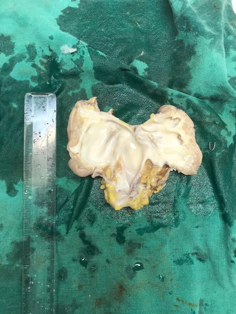
The gross examination of the specimens showed that 20% of the specimen were shrunken, 17% were enlarged and 61% were of normal size. The cortico-medullary junction had lost distinction in 20% of the cases, with thinned out cortex in 30% of the specimens. The dilation of pelvi-calyceal system was observed in 35% of the cases. 56% of the specimen showed one or more cysts. Other findings observed were renal calculi (12%) and gross finding of necrosis and fibrosis (17%). Table 1 shows the distribution of gross appearances of the specimen.
On microscopic examination, the findings were systematically assessed for glomerular, tubular, interstitial and vascular pathology. Table 2 shows the various studied microscopic findings and their distribution. The diagnosis offered after the gross and microscopic examination of the non-neoplastic nephrectomy specimen are tabulated in Table 3.
Table 1
Showing gross appearance of the specimens
Table 2
Showing microscopic findings
Table 3
Showing spectrum of diagnosis of the non-neoplastic nephrectomy specimens
Figure 1
Showing gross features of nephrectomy specimen. Note the lobulations on the surface in cases of chronic pyelonephritis

Figure 2
Showing cut surface of a nephrectomy with dilated pelvi-calyceal system and thinned out cortex

Discussion
Nephrectomy is a common surgical procedure done in cases of untreatable renal pathology including neoplasms. The procedures can be partial nephrectomy or radical nephrectomy. Nephrectomy is also done as a treatment of choice in advanced renal diseases which have caused substantial renal damage deteriorating the renal function totally. The procedure is also done in other cases contributing to loss of renal function like recurrent infection of the pelvicalycal system, calculi, reflux diseases and so on. The studies have shown that the rate at which nephrectomies are done for non neoplastic conditions have gone down over time.4,5,6 In this study, we selected the cases presenting with nephrectomy for non-neoplastic conditions. This was a prospective and retrospective study conducted for duration of three years. A total of 39 cases were obtained.
The year wise incidence of the cases showed that about 81% of the nephrectomies were done for non neoplastic indications. Divyashree et al found the non neoplastic nephrectomy incidence to be 72%.7 Of the total number of cases 21(53%) were males and 18(46%) were females, showing a slight male preponderance. Similar incidence was also found by Divyashree et al but differed from Abdulghafoor S et al who found that 52% were females and 48% were males.8 The age wise incidence of the cases showed that the peak age for nephrectomy was 31-40 yrs for females whereas the males showed two peaks, one at 31-40 yrs and another at 51-60 yrs. Divyashree et al found the peak incidence for both males and females to be at the 3rd decade of life.7
In our study, we found the right sided nephrectomy to be slightly more common than the left side in both male and female cases. About 57% males and 55% females cased had right sided nephrectomy. Gülden Diniz et al in their study found 52% right sided and 47% left sided nephrectomies.9 Divyashree et al found left sided nephrectomies to be slightly more (51%) than right sided (48%).
The commonest clinical indication for nephrectomy was obstructive pathology with calculi at various sites-renal, ureteric, pelvi-ureteric junction. This was followed by hydronephrosis and other miscellaneous conditions including aplastic kidney, cystic disease, reflux nephropathy. Hydronephrosis was by far the most common indication for nephrectomy in both males and females. We found that calculus on radiological examination was by far the most common cause for hydronephrosis. Similar finding was found by Prasanna et al.10 However females showed slightly higher incidence of calculus leading to renal dysfunction. One was a case of aplastic kidney on radiology in which the tissue was removed suspecting neoplasm. Siddappa et al in their study found infective causes for end stage renal disease as the most common indication followed by obstructive causes.11 Abdulghafoor S et al found non functioning kidney as the commonest indication for nephrectomy in their study.
On gross examination of the specimens, we found that majority (61%) of the specimen size were in the normal range. About 20% of them were shrunken and 17% were enlarged in size. Gulden Diniz et al found that 37% of the kidneys were large, 33% were small while 28% were of normal size. On cut surface 20% of the specimens showed loss of cortico-medullary junction and 30% showed thinned cortex. About 56% of the cases showed dilation of pelvi calyceal system. Renal calculi were found in 12% of the cases. Cysts were noted 5% of the cases. Divyashree et al found that 96% of the specimen showed dilatation of pelvi-calyceal system and loss of cortico-medullary junction. They also found that 20% had calculi in the kidneys and 11% showed pus and necrosis. In our study we found areas of necrosis and fibrosis in 17% cases on gross examination.
On microscopic examination we have categorized the description into glomerular, tubular, interstitial and vascular findings. The detailed findings are summerised in table 5
Glomerular findings:
The clinical manifestations leading to nephrectomy in cases of renal failure are predominately the consequence of glomerular lesions. In our study, the predominant finding was glomerular sclerosis in 38% of the specimens and hyalinization of the glomeruli in 23% of the cases. Siddappa S found global glomerular sclerosis as the commonest glomerular findings. The other findings were periglomerular fibrosis and calcification.
Tubular findings
Thyroidisation of the tubules was the most consistent finding with 94% cases showing in variable degrees. Tubules also showed atrophy (23%), dilatation (56%). Few cases showed unremarkable tubules (5%). Siddappa S found tubular atrophy, dilatation and hyaline casts along with thickening of tubular basement membrane as their chief tubular finding.
Interstitial findings
Chronic inflammation was by far the commonest finding with lymphocytes, plasma cells infiltrating the interstitium in all the studied specimens. Areas of interstitial fibrosis were noted in 43% of cases. None of the cases showed xanthogranulomatous changes. Siddappa et al found diffuse interstitial fibrosis and inflammatory infiltrate composed of lymphocytes, plasma cells. Divyashree et al found granulomatous pyelonephritis in 9% of the cases with 6% of them showing features of tuberculosis. One of the cases in our study showed caseous necrosis and well formed epitheloid granuloma with positivity on Zeil-Neilson stain for acid fast bacillus. Another specimen of nephrectomy with aplastic kidney as a radiological diagnosis was one of the nephrectomies under study. We found some fibrous tissue and remnants of seminal vesicles on microscopy.
Vascular findings
Thickening of the vascular wall was noted in more than 50% of the cases and dilatation of the vessel wall in 10% of the specimen on microscopy. However, 38% of the cases showed unremarkable vessel walls.
Spectrum of the non-neoplastic lesions
It was observed that chronic pyelonephritis with end stage kidney disease and hydronephrosis were by far the commonest condition with 88% of the cases. These infection induced poor renal functioning is by far the most common indication for nephrectomy in countries like India.12 The other condition being acute pyelonephritis (2%), chronic interstitial nephritis (10%). One case each of tuberculous pyelonephritis and aplastic kidney were noted. Similar findings were found by Divyashree et al with 84% of the cases being reported as chronic pyelonephritis and 13% as granulomatous pyelonephritis as against 2% in our study. We found no case of xanthogranulomatous pyelonephritis in our study. Divyashree et al found 6% cases as acute pyelonephritis where as we found in 2% of the cases.
Conclusion
This is an elaborate pathological study of non neoplastic nephrectomies in our institution. As majority of the nephrectomy are done for non neoplastic etiology, this study gives an insight of the situation. Our study concludes that chronic pyelonephritis is the commonest non-neoplastic indication for nephrectomy. As this is a treatable condition in the earlier stages, many such nephrectomies can be avoided.


