Introduction
Skin is the largest organ in the body performing multiple functions. More than being a mere protective covering it is a highly sophisticated sensory organ, has endocrine role in synthesising vitamin D.1 Though ubiquitous and simple, it is a heterogeneous organ.2 Histologically it is composed of several cell types that function interdependently and cooperatively.1
The epidermal layer is composed of 90% of keratinocytes and remaining 10% composed by mel anocytes, langerhans cells and merkel cells. Epidermal appendages extend from epidermis to dermis comprising of specialized cells like follicular epithelial cells, sebaceous cells, cells of eccrine and apocrine glands. Different cell types give rise to different types of tumours.3
Skin tumours are generally divided into surface epidermal tumours and tumours of epidermal appendages.2 Epidermis has the capacity to develop into wide variety of lesions though commonly show proliferation of keratinocytes but produce clinically distinct lesions. Adnexal tumours may be solitary or multiple.4,5
These tumours clinically present as papules and nodules. So diagnosis may not be conclusive clinically alone, histopathological confirmation becomes a prime requisite for definite diagnosis. At times it is difficult to interpret because of variety and complexity of histologic nature, complex nomenclature, multiple classifications and conflict of opinion regarding histogenesis.6 Thus clinicopathological correlation is required. Early diagnosis and treatment are necessary for a better cure.
This study was done mainly to evaluate various papular and nodular lesions of skin tumours by histopathological findings.
Materials and Methods
The present study was conducted in the department of pathology, GITAM Institute of Medical Sciences and research, Visakhapatnam. This was a prospective study done during the period of January 2017 to December 2018 i.e. 2 years. Both biopsies and resected specimens were included. The tissues were fixed in 10% formalin and sections were taken. Then they were processed and embedded in paraffin wax. Thin sections of 3-5 microns were made and stained with Haematoxylin and Eosin.
Tumors of epidermis along with melanocytic and adnexae were included in the present study. Mesenchymal tumors, haematological, neural and tumour like lesions including cystic lesions were excluded. The tumours arising from mucocutaneous junction were also excluded from our study.
Results
A total of 46 cases of skin tumours were studied during two years period. Out of 46 cases studied 18 were male (39.14%) and 28(60.86%) female patients with male to female ratio of 2:3 (Table 1).
Tumours of skin are seen in all age groups. The age of the patients ranged from 11 to 68. However most of the tumors are seen in the second and third decade of life (Table 2).
Table 2
Age incidence of individual tumours observed in present study
On histological examination, total benign tumours were 43(93.47%), with only 3 malignant tumours (06.52%). Out of 46 tumours, Epidermal tumours were the most common comprising of 28(60.86%) cases followed by Melanocytic tumours and tumours of Appendageal origin, comprising of 11 (23.9%) and 7(14.2%) cases respectively (Table 3).
Table 3
| Benign | Malignant | Total | |
| Epidermal | 26(56.52%) | 02(4.34%) | 28(60.86%) |
| Melanocytic | 10(21.73%) | 01(2.17%) | 11(23.91%) |
| Appendageal | 07(15.21%) | - | 07(15.21%) |
| Total | 43(93.47%) | 03(6.52%) | 46(100%) |
Distribution of various skin tumours
Tumours from keratinocytic cell of origin were most common in our study. We had a total of 28 cases of tumours of which Squamous papilloma is the most common with about nine cases. It is followed by Achrocordan and Verruca vulgaris. Squamous papillomas were seen in all age groups ranging from 11 years to 61years of age. Most of these patients presented as papillary growth over back and two patients presented over thigh region. Microscopy showed papillomatosis with elongation of rete ridges, hyperkeratosis and acanthosis.
Acrochordans w ere the common tumours next to Squamous papillomas. It is seen in first three decades of life and are not confined to single region but seen in various parts of the body. Histology showed hyperplasia of epidermis with underlying fibrovascular core and loosely arranged collagen. Verruca vulgaris is seen in second to fifth decades of life, which were distributed over head and chest region and one over little finger. All the cases showed hyperkeratosis, acanthosis, papillomatosis and parakeratosis overlying the papillomatous projection along with koilocytic change in the malpighian layer. Verruca plantaris is seen in sixth and seventh decade of life with lesions over the foot region.
Three cases of Seborrheic keratosis were seen in lower part of trunk and scalp region. All are acanthotic type with sheets and columns of basaloid cells and squamoid cells with intervening horn cysts.
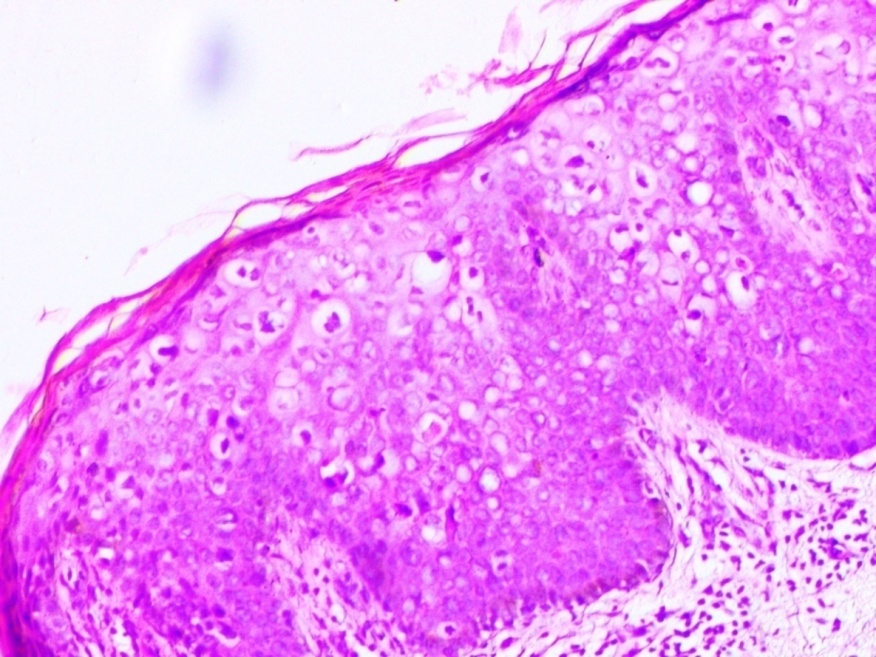
We had a single case of pre malignant condition, Bowen’s disease in a 52 year old female patient who presented as a papular lesion over the abdomen. Microscopy showed hyperproliferative epidermis with marked dysplasia, hyperchromatic nuclei and increased mitotic activity. No breach in to the dermis (Figure 1).
Figure 1
Microscopy of Bowen’s disease showed hyperproliferative epidermis with marked dysplasia, hyperchromatic nuclei and increased mitotic activity. No breach in to dermis

Malignant tumours are very less in our study with only two cases of squamous cell carcinomas which are seen in elderly age group of around 60 years. Of these two one case was seen in a 60 year old female who presented as a ulceroproliferative growth in the elbow, also showed secondary deposits in the Lymphnode. Microscopically both cases were well differentiated
Melanocytic tumours of skin
Intradermal nevus is the most common Melanocytic tumour in our study and is seen in the first three decades of life and the presentation was hyperpigmented papular lesions. Of total eight cases, five cases were seen in the upper trunk and two patients presented over the face. One patient presented as multiple lesions over face and upper back where both biopsies were confirmed as Intradermal nevus. Microscopy showed nevus cells confined to dermis and arranged in nests and cords.
We had a single case of compound nevus on face in a 22 year old male, histology showed nevus cell nests both in the epidermis and dermis. Spitz nevus was seen in a 15 year male on leg which showed large nests of melanocytes between the elongated rete ridges microscopically. A single case of malignant melanoma was seen in 65 year old female over the back. Microscopy showed epithelioid to spindle shaped cells with vesicular nucleus, prominent eosinophilic nucleoli, intra and extracellular melanin pigment (Figure 2).
Adnexal tumours of skin
In the present study there were 7 cases of adnexal tumours and all were benign tumours. Eccrine tumours were the commonest in the study (5 cases) followed by tumours of follicular origin (2 cases).
Two cases of Nodular hidradenoma and Chondroid syringoma were seen in third and sixth decade of life with a single case of Eccrine poroma in a 60 year old female. Microscopy of Nodular hidradenoma showed lobules of varying size s and shapes, consisting of polyhedral cells with round nuclei with slightly basophilic cytoplasm and round cells with clear cytoplasm (Figure 3).
Chondroid syringoma showed cells arranged in solid sheets, cords and ducts intervened by chondromyxoid stroma. Eccrine poroma showed small basaloid cells extending in to dermis in the form of cords and columns.
We have seen a single case of Trichoepithelioma and Pilomatricoma each in second decade of life. Trichoepithelioma histology showed multiple islands and nests of uniform small basoloid cells extending from epidermis to dermis. Pilomatricoma showed sheets of cells with intervening stroma. The cells at the periphery are basoloid with ghost like cells at the centre
Figure 3
Nodular hidradenoma (H&E ×10x) lobules of varying size and shape, consisting of polyhedral cells with round nuclei with slightly basophilic cytoplasm and round cells with clear cytoplasm

Following table (Table 4) shows pattern of distribution of various skin tumours in the present study
Table 4
Distribution of various patterns of skin tumours
Discussion
The tumours of the skin constitute a small but significant proportion of patients. In the present study of 46 cases of skin tumours, there was a female preponderance with a male to female ratio of 1:1.5 and is similar to the study conducted by Namitha et al.7 Benign tumours were more common when compared to malignant tumours, the total number of benign tumours in our study were 43 and our study is comparable to the study done by Rajinder Kaur8 and Narhire V et al.9 where the percentage of benign tumours were 67.27% and 69.4%. But the incidence of benign tumours was very high in our study when compared to other studies. This might be due to difference in the number of cases included, time period of the study and geographical distribution.
Benign tumours were more common when compared to malignant tumours, the total number of benign tumours in our study were 43 and our study is comparable to the study done by Rajinder Kaur8 and Narhire V et al.9 where the percentage of benign tumours were 67.27% and 69.4%. But the incidence of benign tumours was very high in our study when compared to other studies. This might be due to difference in the number of cases included, time period of the study and geographical distribution.
Of all the skin neoplasms, incidence of keratinocytic tumours was high with Squamous Papilloma being the most common keratinocytic tumour.
Malignant tumours are very less in our study with only two cases of squamous cell carcinomas which are seen in elderly age group of around 60 years.
Keratinocytic tumours were followed by Melanocytic tumours and tumours of adnexae. When compared to the other studies done by Shilpa V Uplaonkar10 et al, Shivanand Gundalli et al,11 Vaibhav Bari et al,12 Rajinder Kaur et al8 we had more number of Melanocytic tumours than the tumours of Appendageal origin. Our results are similar to studies done by Samanta M et al13 who had 51.92%, 28.84% and 19.23% of epidermal, Melanocytic and adnexal tumours respectively.
Table 5
Comparision of frequency of various neoplasms of epidermis and epidermal appendages
In the present study we got only seven cases of Appendageal tumours in the two year study period which indicates that adnexal tumours are relatively uncommon. In adnexal tumours, the tumours arising from Eccrine and Apocrine glands were more common and our study can be compared to other studies done by Nair S Patel et al,14 Radhika K et al,15 Samanta M et al13 and Shilpa V Uplaonkar et al.10
Table 6
Comparison of frequency of tumours of epidermal appendages
Similar to the study done by Shilpa V et al10 and Samanta M et al.,13 we too did not get any tumour with sebaceous gland differentiation, may be due to its rare presentation.
Conclusion
Neoplasms of the skin account for a small percentage among all the histopathological lesions. In the present study Keratinocytic tumours are the most common skin tumours of which Squamous papilloma were commonest. These were followed by Melanocytic and Adnexal tumours. Benign tumours were more common when compared to malignant tumours. Maximum tumours were found predominantl in females. Most of the skin tumours present as papules and nodules, at times difficult to diagnose clinically. Hence histopathological examination is must for definite diagnosis and classification of various skin tumours which is helpful for better treatment of the patient.

