- Visibility 85 Views
- Downloads 21 Downloads
- DOI 10.18231/j.ijpo.2019.104
-
CrossMark
- Citation
Clinico-pathological correlation of gastric cancer in a tertiary referral centre
- Author Details:
-
Milind Patil
-
Nilam More *
-
S Solanke
Introduction
Cancer is one of the leading causes of morbidity and mortality worldwide, with approximately 14 million new cases and 8 million cancer related deaths in 2012.[1] Gastric cancer is the fifth most commonly occurring cancer after lung, breast and colorectal cancer. In 2012, of the 952,000 estimated cases, 677,000 cases were from developing countries.
Majority of the patients present with gastric cancer in advanced stage metastasizing to lymph nodes and other organs causing poor prognosis. In Asian countries the intestinal subtype of adenocarcinoma is the commonest histopathological variety. Although surgery is an important part of treatment, long term survival is seen in minority.
This study aims to observe the demographic and clinic-pathological profile of gastric cancer in a tertiary referral centre in Mumbai, India.
Materials and Methods
This was a retrospective study of 81 cases of adenocarcinoma of stomach diagnosed in the Department of Pathology in a tertiary care referral hospital and teaching institute fr om January 2006 to December 2015. Among these cases, 42 specimens and 39 biopsies were studied. The patients were diagnosed on clinical, radiological, endoscopic examination with confirmation by histopathological examination of either endoscopic biopsy or the resected specimen.
Clinical parameters such as age, sex, religion, blood group, dietary habits, addiction, endoscopic findings, clinical features, type of resected specimen, site of lesion, gross findings, histopathological types, associated findings in surrounding mucosa and TNM staging were noted. The resected gastrectomy specimens were examined grossly and tumour location, tumour dimensions, extent of tumour invasion, metastasis to lymph nodes, number of lymph nodes involved, abnormality in surrounding mucosa were recorded.
10% formalin fixed endoscopic biopsy specimens were processed and cut into 4-5 mm thickness and subjected to hematoxylin and eosin (H and E) staining, special stains like periodic acid Schiff (PAS) and alcian blue (AB).
Result
In the period Jan 2006 to Dec 2015, a total of 81 patients presented with gastric cancer. Median age of study population was 60 years. Largest cluster of cases was concentrated in 60-69 years (49%) followed by 50-59 years (27%). Amongst the cases, 50 (62%) were male, 31 (38%) were female, with a sex ratio (male:female) of 1.61:1.
In the study, a considerable population (74.1%) consumed tobacco. The tobacco consumption pattern was found to be more in males (78.0%) as compared with females (67.7%). However, when it came to alcohol consumption, there was significant difference between male (50.0%) and female (3.2%) history. About 75.3% of the population had history of spicy food, while 56.8% had history of salty food. However, consumers of smoked food was comparatively lower (25.9 %). Whereas, only 34.6% of the patients reported consumption of fresh foods. The most common blood group in our study was A (49.4%), followed by O (28.4%).
Abdominal pain was a complaint of all the patients (100%), followed by weight loss (85.2%), nausea (81.5%), anorexia (71.6%), anaemia (56.8%), early satiety (53.1%), mass (38.3%), dysphagia (17.3%) and malena (3.7%). Antrum (46.9%) was the most common site of gastric cancer followed by pylorus (18.5%).
On endoscopy, 32.5% of the cases had ulcerative lesion, followed by diffuse thickening at the distal part seen in 17.5% cases. Histopathologically, according to Lauren’s classification, intestinal type of adenocarcinoma was the most common (59.3%), followed by diffuse type (34.6%) and mixed type (6.2%). Moderately differentiated adenocarcinoma (46.9%) was found to be the most common type followed by poorly differentiated adenocarcinoma (19.8%), signet ring cell adenocarcinoma (16.0%), mucin secreting adenocarcinoma (8.6%), well differentiated adenocarcinoma (7.4%), adenosquamous carcinoma (1.2%). Under TNM staging, 45.2% cases had stage I involvement. 47.6% of the cases had tumour involvement reaching up to serosa i.e. stage II. Very few cases had shown spread of the tumour to the adjacent structure i.e. stage III. Surrounding mucosa showed changes of chronic gastritis in 31.0% of cases and intestinal metaplasia (21.4%) and follicular gastritis (16.7%). However, only 7.1% of cases showed features of atrophic gastritis.
| Age Band | Cases | % |
| 20-29 | 2 | 2.5% |
| 30-39 | 3 | 3.7% |
| 40-49 | 8 | 9.9% |
| 50-59 | 22 | 27.2% |
| 60-69 | 40 | 49.4% |
| 70-79 | 5 | 6.2% |
| 80-89 | 1 | 1.2% |
| Total | 81 | 100.0% |
| Signs and Symptoms | Cases | % |
| Pain in Abdomen | 81 | 100.0% |
| Weight Loss | 69 | 85.2% |
| Nausea/Vomiting | 66 | 81.5% |
| Anorexia | 58 | 71.6% |
| Anaemia | 46 | 56.8% |
| Early Satiety | 43 | 53.1% |
| Mass | 31 | 38.3% |
| Dysphagia | 14 | 17.3% |
| Malena | 3 | 3.7% |
| Site of Lesion | Cases | % |
| Antrum | 38 | 46.9% |
| Fundus | 4 | 4.9% |
| Cardia | 5 | 6.2% |
| Greater Curvature | 6 | 7.4% |
| Lesser Curvature | 13 | 16.0% |
| Pylorus | 15 | 18.5% |
| Total | 81 | 100.0% |
| Types | Total | % |
| Intestinal | 48 | 59.3% |
| Diffuse | 28 | 34.6% |
| Mixed | 5 | 6.2% |
| Total | 81 | 100.0% |


Discussion
Gastric cancer is second most common cancer in male and fifth most common amongst females in East and Central Asia. The incidence of gastric cancer is twice as much in men as in women. Similarly, in our study, the male to female ratio of gastric cancer was 1.61 :1. This was consistent with study conducted by Halder et al.[2] Males have greater exposure to one or more carcinogen and are therefore more susceptible to these factor than women.[3] Further, it is postulated that hormone oestrogen has protective role for gastric carcinoma in females.[4]
It is observed that the incidence of gastric cancer increased with increase in age, peaking at 6th and 5th decade, and then declined in higher age groups.
In our study, the most common blood group among the cases found was A (49.4%), followed by O (28.4%). Wang et al,[5] in their case-control study have found significant relationship between ABO blood groups and gastric cancer, with blood group A having significantly higher risk than that in non-A groups. He concluded that there is an inherited element of susceptibility to cancer of stomach in blood group A that may be partially attributed to an increased risk of H. pylori infection. Jose et al considers that this blood group predisposition is probably related to the nature of mucopolysacharide in gastric secretion and differential susceptibility to damage by environmental carcinogens.
In our study more than half (56.8 %) cases consumed salty food. A high intake of salty food and potentially smoked, cured and pickled foods is thought to be a risk factor. Saghier A et al[6] observed collective evidence from epidemiologic and experimental studies over the past several decades strongly suggests that high intake of salt/salty food is associated with an increased risk of gastric cancer. The acute effects of concentrated salt solutions lead to mucosal damage and its repair is associated with inflammatory changes in human stomach and it also creates condition which enhances H.pylori colonization. Dietary nitrates are found naturally or added during preservation in various foods. Nitrates which are present in fertilizer, soil and water also contributes to dietary nitrates. These nitrates are converted into N-nitroso compounds by gastric acid which are carcinogenic causing increased risk of gastric cancer.
In our study 74.1% of cases had a history of tobacco consumption. Qurieshi et al[7] in their study found that tobacco smoking is an independent risk factor for stomach cancer. Nicotine increases the proliferation and migration of gastric cancer cell by releasing prostaglandins, VEGF and stimulates angiogenesis leading to increase in mediators of cell proliferation, angiogenesis and metastasis and a decrease in apoptosis. Other mechanisms include induction of development of precursor gastric lesions such as gastritis, ulceration and intestinal metaplasia. Smokers tend to have high incidence of H.pylori infection and gastroduodenal infection than non-smokers. However, Rao et al[8] did not find any relation between tobacco use and risk of gastric cancer.
Clinical profile showed that all the patients presented with history of pain in abdomen followed by weight loss (85.2 %). In some studies, abdominal mass in the main presenting complaint in gastric cancer. However, in our study 38.3 % cases presented with mass in abdomen, which was consistent with finding made by Chattopadhyay et al[9] that pain in abdomen was more common than abdominal mass in gastric cancer.
In our study, antrum (46.9%) was the most commonly affected site, followed by pylorus (18.5%), lesser curvature (16.0%) and cardia-fundus (11.1%). This is also supported by report in Indian subcontinent by Halder et al. Chanda et al[10] also observed pyloric antrum as the most common site (65.3%) for gastric carcinomas as pyloric antrum provide favourable environment for H.pylori growth.
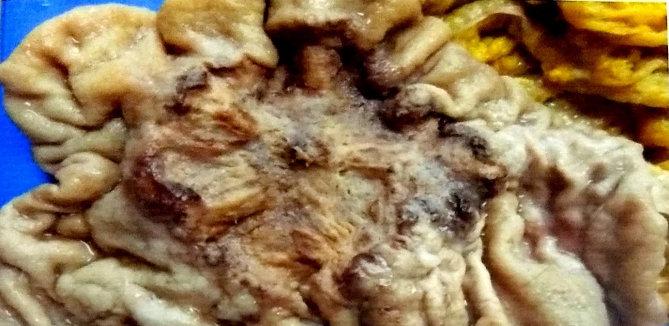
In the present study, among 42 resected specimens, ulcerative growth (46.3 %) was the most common finding on gross examination as well as on endoscopy.
In our study, intestinal adenocarcinoma (59.3%) was most common type, followed by diffuse type (34.6 %). Remaining 6.2 % were of mixed type.
We observed that 46.9 % cases were of moderately differentiated adenocarcinoma, 19.8 % were poorly differentiated, 7.4 % were well differentiated adenocarcinoma of stomach. These findings were consistent with Halder et al where they found 48.6 % and 52.6% of moderately differentiated adenocarcinoma cases respectively.
On TNM staging of gastric cancer, it was observed that 47.6 % cases showed tumour involvement reaching up to serosa, that is Stage II and 38.1% cases showed tumour invading the muscularis propria, that is Stage IB. Very few cases (7.1%) showed spread of the tumour to adjacent structures that is Stage III.
In our study, surrounding mucosa showed presence of chronic gastritis (31.0%), intestinal metaplasia (21.4%), follicular gastritis (16.7%) and atrophic gastritis (7.1%). These findings might be the precursor lesions for gastric cancer. Infection with H. pylori is a major cause of non-atrophic chronic gastritis and is associated with gastric ulcer disease and distal gastric cancer. Intestinal metaplasia have a >10 fold increased risk of developing gastric cancer, which may be even higher in patients with H. pylori infection.
These results re-emphasize that gastric cancer symptoms are non-specific and need an early screening examination.
Conclusion
Present study showed moderately differentiated carcinoma to be the most frequent malignancy. Most patients belonged to 60-69 year age group and had a male predominance. Blood group A was most prevalent and predominant presenting symptom was abdominal pain. More than half of the cases consumed salty food. Majority of cases had history of tobacco consumption, which clearly indicates the role of tobacco in causation of stomach cancer. Weak relation was seen in alcohol consumption. Majority of the cases had ulcerative growth reaching up to serosa, belonged to stage II according to TNM classification. Chronic gastritis may be the precursor lesion for gastric cancer.
Screening of asymptomatic people in a high risk area may be useful in early detection of disease. Efforts to detect cancer early in developing countries would go a long way in reducing the disease burden and improving the outcome.
Source of funding
None.
Conflict of interest
None.
References
- J Ferlay, I Soerjomataram, M Ervik, R Dikshit, S Eser, C Mathers. Cancer Incidence and Mortality Worldwide: IARC CancerBase No. 11 Lyon, France: International Agency for Research on Cancer. 2012. [Google Scholar]
- S K Halder, P K Bhattacharjee, P Bhar, P Bhattacharya, A Pachaury, R R Biswas. Demographic and clinico-pathological profile of carcinoma stomach in a tertiary referral centre of Eastern India. Al Ameen J Med Sci 2012. [Google Scholar]
- L Jose, S Nalappat, V P Sasidharan. A clinico-pathological study of carcinoma stomach. Ind J Pathol Microbiol 1995. [Google Scholar]
- S D Usha, H S Shukla, S Gupta, N C Aryya, S Khanna. A clinicopathological study of carcinoma stomach. Indian J Pathol Microbiol 1988. [Google Scholar]
- Z Wang, L Liu, Ji J .. ABO Blood Group System and Gastric Cancer: A Case-Control Study and Meta-Analysis. Int J Mol Sci 2012. [Google Scholar] [Crossref]
- A A Saghier, J H Kabanja, S Afreen, M Sagar. Gastric cancer: environmental risk factors, treatment and prevention. J Carcinogene Mutagene 2013. [Google Scholar]
- M A Qurieshi, M A Masoodi, S A Kadla, S Z Ahmad, P Gangadharan. Gastric cancer in Kashmir. Asian Pacific J Cancer Prev 2011. [Google Scholar]
- D N Rao, B Ganesh, K A Dinshaw, K M Mohandas. A case-control study of stomach cancer in Mumbai, India. Int J Cancer 2002. [Google Scholar]
- S D Chattopadhyay, R K Singhamahapatra, R S Biswas, T K Sengupta, A Bandopadhyay, N C Nath. Prevalence of carcinoma stomach in a tertiary referral centre in Eastern India and its correlation with endoscopic findings. J Indian Med Assoc 2011. [Google Scholar]
- N Chanda, A R Khan, M Romana, S Lateef. Histopathology of Gastric Cancer in Kashmir-A Five Year Retrospective Analysis. 2007. [Google Scholar]
